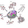
Cell Synchronization is a process by which
cells at different stages of the cell cycle in a culture are brought to the
same phase.[1] "Cell synchrony" is required to study the progression
of cells through the cell cycle. The types of synchronizations are broadly
categorized into two groups: "Physical Fractionation" and
"Chemical Blockade."
1.
Cell separation by physical means:
Physical fractionation or cell separation
techniques, based on the following characteristics are in use: Cell density; Cell
size; Affinity of antibodies on cell surface epitopes; Light scatter or
fluorescent emission by labeled cells.
The two commonly used techniques are:
(1)Centrifugal
separation
The physical characteristics — cell size
and sedimentation velocity — are operative in the technique of centrifugal
elutriation. Centrifugal elutriator (from Beckman) is an advanced device for
increasing the sedimentation rate so that the yield and resolution of cells is
better. The cell separation is carried out in a specially designed centrifuge
and rotor.
(2)Fluorescence-activated
cell sorting
Fluorescence-activated cell sorting (FACS)
is a technique for sorting out the cells based on the differences that can be
detected by light scatter (eg. cell size) or fluorescence emission (by
penetrated DNA, RNA, proteins, antigens). The procedure involves passing of a
single stream of cells through a laser beam so that the scattered light from
the cells can be detected and recorded. There are two instruments in use based
on its principle:
a) Flow cytometer
b) Fluorescence-activated cell sorter
2.
Cell separation by chemical blockade
The cells can be separated by blocking
metabolic reactions.[2] Two types of metabolic blockades are in use:
(1)Inhibition
of DNA synthesis
During the S phase of cell cycle, DNA
synthesis can be inhibited by using inhibitors such as thymidine, aminopterin,
hydroxyurea and cytosine arabinoside. The effects of these inhibitors are
variable. The cell cycle is predominantly blocked in S phase that results in
viable cells.
(2)Nutritional
deprivation
Elimination of serum from the culture
medium for about 24 hours results in the accumulation of cells at G1 phase.
This effect of nutritional deprivation can be restored by their addition by
which time the cell synchrony occurs.
Ref:
1.Merrill
GF (1998). "Cell synchronization.". Methods in Cell Biology 57:
229–249. doi:10.1016/S0091-679X(08)61582-4. PMID 9648108.
2.Davis
PK, Ho A, Dowdy SF (2001). "Biological methods for cell-cycle
synchronization of mammalian cells". BioTechniques 30 (6): 1322–1331. PMID
11414226.
Attached:
Protocol 1
Metaphase Block
for Cell Synchronization
Posted on Monday, October 27, 2003
Description
Metaphase Block for Cell Synchronization
Procedure
1. Remove the medium from an exponentially-growing cell culture and rinse it
with 10 ml of PBS. (See Hint #1)
2. Replace the PBS with Complete Medium with Nocodazole and allow the culture
to incubate at 37°C for 12 hr.
3. Shake off loosely attached, rounded mitotic cells by gently knocking the
plate and pipetting medium over the cell layer a few times.
4. Collect the cell-containing medium and place it in a sterile 50 ml
polypropylene tube and pellet the cells by centrifugation at 1000 X g for 10
min. Remove the supernatant (the cell pellet may be small) and resuspend the
cells in 15 ml of PBS.
5. Repellet the cells at 1000 X g for 10 min and remove the PBS. Resuspend the
cells in complete medium and proceed with experiment or return the cells to cell
culture flask and incubate at 37°C.
Recipes
PBS pH 7.2
2.7 mM KCl
4.3 mM Sodium Phosphate Dibasic (Na2HPO4)
1.8 mM Potassium Phosphate Monobasic (KH2PO4)
137 mM NaCl
Complete Medium 100 Units/ml Penicillin
100 ìg/ml Streptomycin
DMEM
10% (v/v) Calf Serum
Nocodazole Stock (10,000 X) 6 g/ml in DMSO
(CAUTION See Hint #2)
Complete Medium with Nocodazole 100 Units/ml Penicillin
Dulbecco's Modified Eagle's Medium (DMEM)
100 ìg/ml Streptomycin
10% (v/v) Calf Serum
600 ng/ml Nocodazole (diluted from Nocodazole stock)
Supplies
Tips
1. Different cell lines use different media, thus "complete medium"
may be different for your cell line than what is described here.
Protocol 2
Cell synchronization using a double thymidine block
1. Cells are grown to a subconfluent density (mid-log phase) in serum-rich
medium.
2. Thymidine is
added to 2 mM final and incubated for 16 hours for HEp-2, 19 h for HeLa.
3. Cells are
washed three times with PBS on plates and refed with fresh serum-rich medium.
4. Incubate for 10
hours before adding 2 mM thymidine.
5. Incubate for 14
hours (17 for HeLa) and wash as described in step 3, refeed, and begin time
points.
Note: Conditions
for synchronization of different cell lines may vary (e.g. time of incubation).
From:http://elledgelab.bwh.harvard.edu/protocols/mamCell/cellsyn.html