利用组织成像质谱技术分析伊立替康在肝脏和人类肿瘤中的分布
前言:
Purpose: To determine irinotecan metabolites and their distribution in human tumors directly from tissue by MALDI Imaging mass spectrometry. To establish MALDI ion trap technology as ideally suited for direct tissue analysis of drugs in tissue.
Methods: Nude mice with implanted human head and neck tumors were treated with either irinotecan, methylselenocysteine (MSC), both drugs combination, or non-treated (control). Frozen tumors and livers were sliced to ~12μm thick and thaw-mounted on to non-conductive glass slides. Irinotecan metabolites were investigated by Single Reaction Monitoring or CRM with an LTQ XLTM coupled to a MALDI source.
Results: Metabolites previously identified in human urine samples were detected directly off tissue in all irinotecan treated tissues and those with both irinotecan plus MSC. SN-38-G, a known irinotecan metabolite, seems more uniformly distributed in the tumor treated with irinotecan plus MSC.
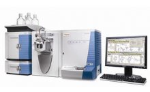
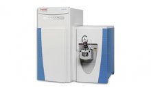
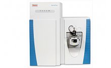
仪器:
结论:
- Most known irinotecan metabolites were identified by MS/MS and confirmed by MS3 using the LTQ XL with MALDI source directly from drug-treated tumor samples.
- The irinotecan metabolite SN-38-G seemed more uniformly distributed in FaDu tumors treated with both MSC and irinotecan than in those treated with irinotecan alone, where the analyte appears clumped in regions (MS2 and MS3 data).
- Rastering a commercial laser with beam of ~100 μm at a spacing of 50 μm does improve the resolution of the 2-dimensional image5 (Fig. 4 a, c).
- Given the complexity of tissue samples, MS3 is truly needed in MALDI tissue imaging to filter interfering and isobaric species that are part of the baseline chemical noise and isolated during MS2.
- Future work using MALDI tissue imaging will compare the results presented here in highly vascularized FaDu tumors with avascular tumors such as A253.


