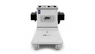
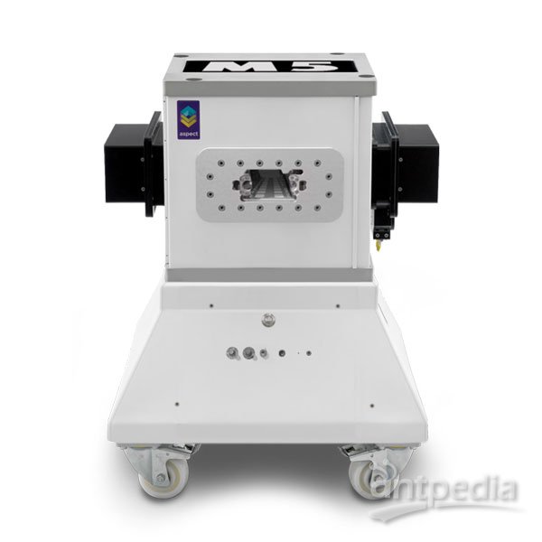
活体GFP绿色荧光成像系统
| 系统提供动物活体绿色荧光蛋白的实时观察与成像等一系列的荧光检测。能够应用在像深度肿瘤,大动物等活体肿瘤追踪观察成像研究。 该设备是一个高灵敏度的图像成像工作系统,主要利用特定波长的激光进行激发后,通过高灵敏度的致冷CCD进行实时检测后,获得所需的各类 特性的图像,有利于进一步的分析作用 。 采用高灵敏度CCD成像系统, 通过长时间积分功能,可拍摄人的肉眼不可见荧光信号,实现成像。 Imaged using a Zoom Lens with 515nm viewing filter and Macro-ImagerCamera, 470nm excitation using the Illumatool TLS LT-9500 same mouseboth images . Mouse Images Courtesy of AntiCancer , Inc |
|
|
全文下载 (PDF, 838 KB)
Time-dependent whole-body fluorescence tomography of probe bio-distributions in mice
We present a fast scanning fluorescence optical tomography system for imaging the kinetics of probe distributions through out the whole body of small animals. Configured in a plane parallel geometry, the system scans a source laser using a galvanometer mirror pair (τswitch~1ms) over flexible source patterns, and detects excitation and emission light using a high frame rate low noise, 5 MHz electron multiplied charge-coupled device (EMCCD) camera. Phantom studies were used to evaluate resolution, linearity, and sensitivity. Time dependent (δt=2.2 min.) in vivo imaging of mice was performed following injections of a fluorescing probe (indocyanine green). The capability to detect differences in probe delivery route was demonstrated by comparing an intravenous injection, versus an injection into a fat pocket (retro orbital injection). Feasibility of imaging the distribution of tumor-targeted molecular probes was demonstrated by imaging a breast tumor-specific near infrared polypeptide in MDA MB 361 tumor bearing nude mice. A tomography scan, at 24 hour post injection, revealed preferential uptake in the tumor relative to surrounding tissue.


