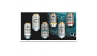
Microscopy with Oil Immersion
Microscopy with Oil Immersion
Principle
When light passes from a material of one refractive index to material of another, as from glass to air or from air to glass, it bends. Light of different wavelengths bends at different angles, so that as objects are magnified the images become less and less distinct. This loss of resolution becomes very apparent at magnifications of above 400x or so. In fact, as you will see later, even at 400x the images of very small objects are badly distorted.
Placing a drop of oil with the same refractive index as glass between the cover slip and objective lens eliminates two refractive surfaces and considerably enhances resolution, so that magnifications of 1000x or greater can be achieved.
To use an oil immersion lens, first focus on the area of specimen to be observed with the high dry (400x) lens. Pace a drop of immersion oil on the cover slip over that area, and very carefully swing the oil immersion lens into place. Focus carefully, preferably by observing the lens itself while bringing it as close to the cover slip as possible, then focusing by moving the lens away from the specimen. When in focus the lens nearly touches the cover slip. The focal plane is so narrow that it is very easy to focus right past it. If you are focusing toward the specimen, you can drive the lens right into it.
When to use oil immersion lenses
Use an oil immersion lens when you have a fixed (dead - not moving) specimen that is no thicker than a few micrometers. Even then, use it only when the structures you wish to view are quite small - one or two micrometers in dimension. Oil immersion is essential for viewing individual bacteria or details of the striations of skeletal muscle. It is nearly impossible to view living, motile protists at a magnification of 1000x, except for the very smallest and slowest.
A disadvantage of oil immersion viewing is that the oil must stay in contact, and oil is viscous. A wet mount must be very secure to use oil. Oil immersion lenses are used only with oil, and oil can't be used with dry lenses, such as your 400x lens. Lenses of high magnification must be brought very close to the specimen to focus and the focal plane is very shallow, so focusing can be difficult. Oil distorts images seen with dry lenses, so once you place oil on a slide it must be cleaned off thoroughly before using the high dry lens again. Oil on non-oil lenses will distort viewing and possibly damage the coatings.
Exercise: examination of stained bacteria
Bacteria can readily be seen even with the scanning lens, provided sufficient contrast can be provided. Dark field microscopy is best for detecting motile bacteria at low power, for example. When bacteria are heat-fixed and stained they tend to clump together. At high dry power individual cells often can't be detected because the space between cell walls is within the limits of resolution of the microscope due to diffraction. To properly view stained bacteria it is necessary to use oil immersion microscopy.
Obtain a slide of gram stained bacteria. Each slide is etched on the lower surface, with the heat-killed and stained bacteria on the opposite (upper) surface. Mount the slide with smear up and focus at low power on the etching. Move the slide so that the lens is over the smear itself. Now move the objective away from the slide until the upper surface comes into focus.
A bacterial smear at low power looks like a patch of dirt. Focus on the mess, first at 40x then at 100x and finally 400x, all in bright field mode. Note that you can see some detail at 400x, but the shapes and colors of the bacteria are somewhat distorted.
Move the 400x lens out of the way, place a drop of immersion oil directly on the smear where the objective was, and swing the oil immersion lens in place. Move the fine focus up and down slightly to ensure that the lens is in contact with the oil. Now watch the end of the objective and bring it as close to the slide surface as you can without touching it. Note in which direction you must focus to move the objective away from the slide, look in the eyepieces, and slowly rotate the fine focus control until the image is focused.
Most bacterial species are rod-shaped or round (cocci), although some are curved, spiral-shaped, or irregularly shaped. The gram stain leaves some cell types pink (Gram negative) and others dark blue (Gram positive) depending on cell wall characteristics. Gram stain results are a major criterion for identification of species. Note how much more clear the image is at 1000x with oil than it was at 400x without oil


