Product Details
The RMP1-14 monoclonal antibody reacts with mouse PD-1 (programmed death-1) also known as CD279. PD-1 is a 50-55 kDa cell surface receptor encoded by the Pdcd1 gene that belongs to the CD28 family of the Ig superfamily. PD-1 is transiently expressed on CD4 and CD8 thymocytes as well as activated T and B lymphocytes and myeloid cells. PD-1 expression declines after successful elimination of antigen. Additionally, Pdcd1 mRNA is expressed in developing B lymphocytes during the pro-B-cell stage. PD-1’s structure includes a ITIM (immunoreceptor tyrosine-based inhibitory motif) suggesting that PD-1 negatively regulates TCR signals. PD-1 signals via binding its two ligands, PD-L1 and PD-L2 both members of the B7 family. Upon ligand binding, PD-1 signaling inhibits T-cell activation, leading to reduced proliferation, cytokine production, and T-cell death. Additionally, PD-1 is known to play key roles in peripheral tolerance and prevention of autoimmune disease in mice as PD-1 knockout animals show dilated cardiomyopathy, splenomegaly, and loss of peripheral tolerance. Induced PD-L1 expression is common in many tumors including squamous cell carcinoma, colon adenocarcinoma, and breast adenocarcinoma. PD-L1 overexpression results in increased resistance of tumor cells to CD8 T cell mediated lysis. In mouse models of melanoma, tumor growth can be transiently arrested via treatment with antibodies which block the interaction between PD-L1 and its receptor PD-1. For these reasons anti-PD-1 mediated immunotherapies are currently being explored as cancer treatments. Like the J43 antibody the RMP1-14 antibody has been shown to block the binding of both mouse PD-L1-Ig and mouse PD-L2-Ig to PD-1.

RMP1-14单克隆抗体与小鼠PD-1(程序性死亡-1)反应,也称为CD279。PD-1是由Pdcd50基因编码的55-1 kDa细胞表面受体,属于Ig超家族的CD28家族。PD-1在CD4和CD8胸腺细胞以及活化的T和B淋巴细胞和骨髓细胞上短暂表达。PD-1在成功消除抗原后表达下降。此外,Pdcd1 mRNA在前B细胞阶段的发育B淋巴细胞中表达。PD-1的结构包括ITIM(基于免疫受体酪氨酸的抑制基序),表明PD-1负调节TCR信号。PD-1通过结合其两个配体PD-L1和PD-L2来发出信号,PD-L7和PD-L1都是B1家族的成员。配体结合后,PD-1信号传导抑制T细胞活化,导致增殖减少,细胞因子产生和T细胞死亡。
此外,已知PD-1在小鼠外周耐受和预防自身免疫性疾病中起关键作用,因为PD-1基因敲除动物表现出扩张型心肌病,脾肿大和外周耐受性丧失。诱导PD-L8表达在许多肿瘤中很常见,包括鳞状细胞癌、结肠腺癌和乳腺腺癌。PD-L1过表达导致肿瘤细胞对CD1 T细胞介导的裂解的耐药性增加。在黑色素瘤小鼠模型中,肿瘤生长可以通过阻断PD-L1与其受体PD-43之间相互作用的抗体治疗来暂时阻止。由于这些原因,目前正在探索抗PD-1介导的免疫疗法作为癌症治疗方法。与J14抗体一样,RMP1-2抗体已被证明可以阻断小鼠PD-L1-Ig和小鼠PD-L<>-Ig与PD-<>的结合。
艾美捷体内小鼠PD-1抗体#BE0146详细信息:
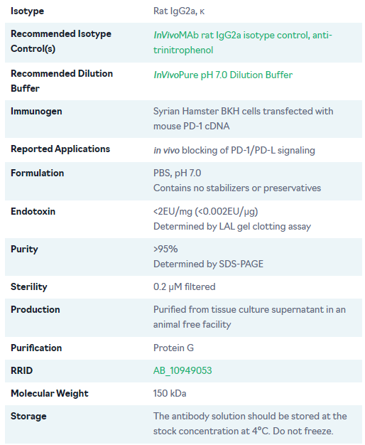
同种型 大鼠IgG2a, κ
推荐的同种型对照 体内MAb大鼠IgG2a同种型对照,抗三硝基苯酚
推荐的稀释缓冲液 体内纯 pH 7.0 稀释缓冲液
免疫原 用小鼠PD-1 cDNA转染的叙利亚仓鼠BKH细胞
应用程序 PD-1/PD-L信号传导的体内阻断
配方 PBS,pH 7.0
不含稳定剂或防腐剂
内毒素 <2EU/mg (<0.002EU/μg)
通过LAL凝胶凝血测定法测定
纯度 >95%
由 SDS-PAGE 决定
无 菌 0.2 μM 过滤
生产 从无动物设施中的组织培养上清液中纯化
纯化 蛋白 G
里德 AB_10949053
分子量 150 千达
存储 抗体溶液应以4°C的储备浓度储存。 不要冻结。
体内小鼠PD-1抗体部分文献参考:
in vivo blocking of PD-1/PD-L signaling
Ngiow, S. F., et al. (2015). "A Threshold Level of Intratumor CD8+ T-cell PD1 Expression Dictates Therapeutic Response to Anti-PD1" Cancer Res 75(18): 3800-3811.
in vivo blocking of PD-1/PD-L signaling
Evans, E. E., et al. (2015). "Antibody Blockade of Semaphorin 4D Promotes Immune Infiltration into Tumor and Enhances Response to Other Immunomodulatory Therapies" Cancer Immunol Res 3(6): 689-701.
in vivo blocking of PD-1/PD-L signaling
Zelenay, S., et al. (2015). "Cyclooxygenase-Dependent Tumor Growth through Evasion of Immunity" Cell 162(6): 1257-1270.
来源:https://www.amyjet.com/featured/mtdna-target.shtml