| INTRODUCTION |
This protocol describes the histological preparation of embryonic and adult zebrafish eyes. The methods described here can be easily adapted for use on other zebrafish tissues.
|
| RELATED INFORMATION |
These methods have been employed in studies on morphological development of the zebrafish retina (Branchek and Bremiller 1984, Schmitt and Dowling 1999).
|
MATERIALS
|
Reagents
|
0.01 M phosphate buffered saline (PBS) (pH 7.4) (Sigma)
DPX mountant (Electron Microscopy Sciences, 13512)
Embedding resin for zebrafish eyes
ReagentAmount to addEpon 81225 mLAraldite 50220 mL2-Dodecenylsuccinic acid anhydride (DDSA)60 mLDMP-301.1 mLSolution must be mixed thoroughly. Store at -20°C.
Ethanol (50%, 70%, 80%, 90%, and 100%)
Fixation solution for zebrafish eyes
1% (w/v) paraformaldehyde (PFA)
3% (w/v) sucrose
2.5% (v/v) glutaraldehyde
0.2 M phosphate buffer (pH 7.4)
This solution can be stored at 4°C for 2 wk.
Histology stain
|
Equipment
|
Coverslips, No. 1 thickness
Embedding mold
Fume hood
Heating block
Knives, glass
Microscope, light
Microtome
Needles (25-30 gauge)
Oven, preset to 60°C
Razor
Scalpel
Slides, gelatin coated or positively charged (e.g., ESCO Superfrost Plus, Erie Scientific)
Spatula, wooden
Stereomicroscope
Transfer pipettes, disposable
Tubes (1.5 mL), microcentrifuge
Tweezers
Vials (15 mL), conical
|
METHOD
|
FixationCollect and fix surgically removed adult eyes or entire embryonic or larval fish in fixation solution:
To dissect and fix adult eyes, euthanize fish in tricaine. Use a scalpel and fine tweezers to remove the eyes, and puncture the corneas with a 25- to 30-gauge needle. Place the eyes in a 15-mL conical vial with 3-4 mL of fixation solution. Fix at 4°C for 2-3 d. If desired, place one eye in fixation solution for histology, and process the contralateral eye for immunohistochemistry in 4% PFA as described in Immunohistochemistry on Cryosections from Embryonic and Adult Zebrafish Eyes (Uribe and Gross 2007).
To fix larval fish, place the entire fish in a 1.5-mL microcentrifuge tube containing fixation solution. Fix overnight at 4°C or at room temperature for 4-6 h.
Remove the fixation solution and wash the fixed samples in 0.01 M PBS:
Wash adult samples three times for 15 min per wash.
Wash larval samples three times for 5 min per wash.
Fix the samples in 1% osmium tetroxide at 4°C:
Fix adult eyes for 2-3 h.
Fix larval samples for 60-90 min. Avoid light during fixation.
Remove the osmium tetroxide and wash samples three times in 0.01 M PBS for 5 min per wash.
Osmium tetroxide is toxic and should be disposed of with hazardous waste.
Remove the PBS and proceed with the following dehydration series:
Care should be taken with propylene oxide as it is highly volatile; propylene oxide incubations and the subsequent steps should be performed in a hood until the samples are in embedding resin and transferred to an incubator (Step 9).
Thaw embedding resin during the second propylene oxide incubation. The resin is highly toxic until it has polymerized. If possible, devote specific pieces of equipment to resin-related work (i.e., 60°C incubator, heating/stirring block, stereomicroscope).
50% ethanol for 5 min
70% ethanol for 10 min
80% ethanol for 15 min
90% ethanol for 20 min
100% ethanol for 15 min
100% ethanol for 15 min
100% propylene oxide for 10 min
100% propylene oxide for 10 min
Carefully mix equal amounts of embedding resin and 100% propylene oxide in a 15-mL conical tube. Begin resin infiltration with 50% propylene oxide and 50% resin for 1 h or longer.
Samples may be left for 5-6 h.
Remove the 50/50 embedding resin/propylene oxide mix and add 100% embedding resin to the samples. Leave the samples overnight at room temperature in the hood with caps open, to allow for propylene oxide evaporation.
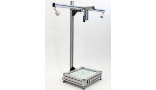
The next day, remove the samples and place them in fresh embedding resin in an embedding mold. Under a stereomicroscope, align the samples using a small needle:
Place one to five embryos at the end of a single well, dorsal side up, and align as closely as possible to each other (Fig. 1A ).

Figure 1. (A) Alignment of 9-dpf (days post-fertilization) embryos in a histology mold. (B) Alignment of an adult eye in a histology mold.
Place a single adult eye, lens side up, in a well (Fig. 1B).
It is helpful to place a small piece of paper with an identifying name or code into each well before adding the resin and samples.
Bake in a 60°C oven for 2-3 d. Baking time depends on the batch of resin. Once the resin has polymerized, the blocks can be stored indefinitely at room temperature until ready for sectioning.
Sectioning
Trim the block with a razor by cutting excessive plastic (polymerized resin) away from the samples. Align the specimen in the microtome block holder to provide the correct sectioning angle.
Using a glass knife and microtome, cut thick sections (5-10 µm) until the desired location or depth is reached. To determine location, check the sections often using a light microscope.
Cut semithin histological sections, 1-1.5-µm thick. Using a wooden spatula (the shaved end of an applicator tip), collect the sections and place them on a droplet of H2O on a gelatin-coated slide.
Place the slide on a heating block to evaporate the H2O droplet and allow the specimen to adhere to the slide.
Stain the sections with histology stain for 30 sec to 3 min. Stain time varies with the batch of stain and batch of resin.
Rinse the sections with H2O and check the samples on a microscope for the desired level of staining.
If the sections have not absorbed enough stain, repeat Step 14.
If samples are appropriately stained, air-dry for 10-15 min.
Using a disposable transfer pipette, add three to four small drops of DPX mountant.
The use of disposable pipettes eliminates the risk of contaminating more expensive pipettes with the mountant.
Place a No. 1 coverslip onto each sample and let the DPX mountant harden overnight.
Image on a light microscope (Fig. 2 , Fig. 3 ).

Figure 2. Transverse histological section of 5-dpf zebrafish eye stained with histology stain (methylene blue and azure B). The dorsal side is up.

Figure 3. Transverse histological section of an 18-mo-old zebrafish eye stained with histology stain (methylene blue and azure B). The ventral retina is shown.
|
ACKNOWLEDGMENTS
|
This protocol describes methods originally developed in John E. Dowling's laboratory at Harvard University. Work in J.M.G.'s laboratory is supported by the Knights Templar Eye Foundation, the American Health Assistance Foundation Macular Degeneration Research Program, and the Retina Research Foundation. We thank Polly Harvey for technical assistance.
|
REFERENCES
|
Branchek, T. and Bremiller, R. 1984. The development of photoreceptors in the zebrafish, Brachydanio rerio. I. Structure. J. Comp. Neurol. 224: 107-115.
Schmitt, E.A. and Dowling, J.E. 1999. Early retinal development in the zebrafish, Danio rerio: Light and electron microscopic analyses. J. Comp. Neurol. 404: 515-536.
Uribe, R.A. and Gross, J.M. 2007. Immunohistochemistry on cryosections from embryonic and adult zebrafish eyes. CSH Protocols doi: 10.1101/pdb.prot4779.
|
Copyright © 2007 by Cold Spring Harbor Laboratory Press. Online ISSN: 1559-6095 Terms of Service All rights reserved. Anyone using the procedures outlined in these protocols does so at their own risk. Cold Spring Harbor Laboratory makes no representations or warranties with respect to the material set forth in these protocols and has no liability in connection with their use. Materials used in these protocols may be considered hazardous and should be used with caution. For a full listing of cautions regarding these material, please consult:
CSH Protocols; 2007; doi:10.1101/pdb.prot4846
http://www.cshprotocols.org/cgi/content/full/2007/18/pdb.prot4846
|