A simple, rapid procedure for the isolation of DNA for PCR
The polymerase chain reaction (PCR) is a method for amplifying specific segments of DNA defined by the small primers used to start the reaction. Using arbitrarily chosen 10-base primers, one can generate "random amplified polymorphic DNA" (RAPD) markers (Williams et al. 1991 Nucl. Acids Res. 18:6531-6535). These DNA fragments, separated by electrophoresis in an agarose gel, can be used as markers for studying genetic variation within and among fungal populations. A rapid and simple procedure for isolating fungal DNA from multiple isolates is needed if this technique is to be useful for diagnosis and screening of natural populations. We have developed a method for isolating DNA without grinding from Fusarium cultures grown on agar slants using a modification of the SDS miniprep method of Lee et al. (1988 Fungal Genetics Newsletter 35:23-24).
1. Place a cube (0.5 cc) of mycelia-covered agar from a 5 day-old slant in a 1.5 ml microcentrifuge tube.
2. Fill tube with liquid nitrogen and let it evaporate. Repeat. No grinding is necessary.
3. Immediately add 500 ul of 65 C lysis buffer (50 mM Tris pH 8, 50 mM EDTA, 3 SDS, 1 BME, and 0.1 mg/ml Proteinase K). Vortex tube and place in 65 C water bath for 1 hr. Vortex after 30 min and 60 min.
4. Extract with 500 ul phenol. Spin tube 5 min at 8000 rpm in microcentrifuge to separate phases. Remove 450 ul of the aqueous phase.
5. Extract with 450 ul buffered phenol. Spin. Remove 400 ul of the aqueous phase. 6. Extract with 400 ul chloroform:isoamyl alcohol::24:1. Spin. Remove 350 ul of the aqueous phase.
7. Add 50 ul 7.5 M ammonium acetate. Gently mix. Add 880 ul 95 ethanol. Invert to mix. Place in -20 C freezer 30 min to overnight.
8. Spin down DNA pellet for 20 min at 13,000 rpm. Rinse pellet in 70 ethanol. Dry pellet and resuspend in 20 ul TE (10 mM Tris, 1 mM EDTA, pH 8).
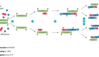
DNA was prepared from isolates grown on complete and minimal slants (Correll et al. 1987 Phytopathology 77:1640-1646). We obtained enough DNA from each isolate for 20 reactions. We used 1 ul of DNA for PCR with our primer, designated ECORI (5''- ATGAATTCGC-3''). To each reaction tube on ice was added 42 ul sterile, glass-distilled and deionized water; 5 ul 10X buffer from Promega (500 mM KCl, 100 mM Tris-HCl pH 9, 15 mM MgCl, 0.1 gelatin w/v, 1 Triton X-100), 1 ul of 50 uM primer, 1 ul of 1 mM dNTP''s and 1 ul DNA solution. Tubes were boiled for 2.5 min. Tubes were returned to ice, then 1 unit of Taq polymerase (Promega) was added. Tubes were spun for 5 sec in a 4 C microfuge and then 100 ul mineral oil was added. Tubes were placed in a PTC-100 programmable thermal controller from M.J. Research with the following program: Step 1. 92 C 30 sec. Step 2. 35 C 1 min. Step 3. Slope 35 to 72 at 1 degree C every 8 sec. Step 4. 72 C 2 min. Step 5. cycle to Step 1. 45 times. Step 6. 72 C 7 min. and Step 7. end. 20 ul of each reaction mix was loaded into a 1.3 agarose gel with TBE buffer. After 2.0 hours at 56 V, the gel was stained with ethidium bromide and photographed. The resulting patterns. of amplified DNA fragments are listed in Table 1.
Table 1. RAPD marker patterns for Gibberella fujikuroi isolates using the ECORI primer
Strain    DNA fragments,  kb +/- SE
A2910     1.661 +/- .044, 0.597 +/- .015
A2911     1.698 +/- .098, 0.561 +/- .031
A3957     1.678 +/- .032, 0.639 +/- .015
X3974     1.661 +/- .044
Acknowledgements: Contribution 91-404-J, Kansas Agricultural Experiment Station, Kansas State University, Manhattan. Supported by a grant from the Kansas State Board of Agriculture (Corn Commission and Grain Sorghum Commission), by the Kansas Agricultural Experiment State Hatch Project 547, and by the Sorghum/Millet Collaborative Research Support Program (INTSORMIL) AID/DAN-1254-G-00-0021-00 from the Agency for International Development, Washington, DC.