Handling Fresh Hepatocytes in Suspension
实验概要
Important—Please read!
GIBCO® Fresh Hepatocytes are shipped in cold preservation medium designed to keep cells viable at 4°C. This shipping medium is not suitable for maintenance of hepatocytes in culture and should be exchanged with new medium, using the following instructions, immediately upon receipt.
Precautions
Treat all hepatocytes as potentially hazardous material.
Wipe surfaces, and exteriors of bottles and hepatocyte containers with 70% isopropanol.
Perform all operations using aseptic handling technique in a sterile environment.
主要试剂
Dexamethasone, 10 mM in DMSO solution, 1 x 250 μL (supplied in excess)
Maintenance Cocktail solution, 1 x 20 mL, containing:
2.5 mL penicillin-streptomycin, 10,000 U/mL
5.0 mL ITS (insulin, transferrin, selenium complex, BSA, linoleic acid)
5.0 mL GlutaMAX™, 100X
7.5 mL HEPES, pH 7.4; 1M
实验步骤
1. Centrifuge and resuspend hepatocytes
1) Remove tube(s) of hepatocytes from shipping container; record lot number.
2) Slowly invert tube a few times to mix cell suspension.
3) Centrifuge hepatocytes at 4°C :
Human hepatocytes - 100 x g for 10 min
Animal hepatocytes - 76 x g for 6 min
4) Aspirate supernatant carefully, leaving approximately 1 mL of medium in tube.
5) Gently resuspend pellet in either incubation or plating media - target 1 million cells/mL unless experiment requires higher concentration of cells.
2. Determine viability and viable cell yield by Trypan Blue exclusion count
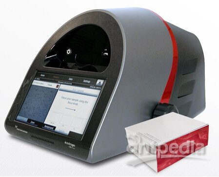
1) Mix 400 μL buffer (PBS or similar) with 50 μL 0.4% Trypan Blue stain (Invitrogen Cat. No. T10282) in a 1.5 mL microcentrifuge tube (this is the cell counting vial).
2) Before adding a portion of the hepatocyte suspension to the counting vial, gently invert the tube of hepatocytes several times to ensure a homogeneous solution. This step is crucial to obtaining an accurate cell count.
3) Using a wide-bore tip, pipette 50 μL of the hepatocyte suspension into the cell counting vial by drawing the solution into the pipette tip once and depressing the contents into the cell counting vial. Do not rinse out the tip. This results in a 10-fold dilution of the original cell suspension.
4) Gently flick/tap the counting vial to ensure a homogeneous mixture. Do not shake or agitate vigorously, as this will cause cell death and result in an inaccurate viability determination.
5) Wait one minute, gently flick/tap the counting vial again to mix, and then slowly pipette 10 μL of the mixture into the V-shaped groove on one side of the hemocytometer. Capillary action will pull the cell suspension into the hemacytometer.
6) Before counting, quickly scan the quadrants under the microscope to ensure the cells are evenly distributed. To ensure an accurate cell count, aim to have approximately 80 to 140 cells per quadrant. Make note if you change dilution factors, and reload if the distribution appears uneven or contains bubbles.
7) Immediately count the viable hepatocytes (clear or yellow color) and dead hepatocytes (blue color) in the four outer quadrants of the hemocytometer.
8) Determine viability of hepatocytes (live cells/total cells x 100).
9) Determine total cell density (cells/mL = average number of live cells x 10,000 x 10). Where 10,000 is the surface area of the hemacytometer and 10 is the dilution factor.
10) To use hepatocytes in suspension assays, adjust cell density to 0.5-2.0 x 10⁶ viable cells/mL as experiment requires.
11) To use hepatocytes in a plated format, adjust viable cell density according to plate format and species. See tables per species per plate format for recommendations. Note: It is recommended to do a visual density check to ensure your seeding density is accurate.