Cell丨施一公组完成酵母剪接体结构最后拼图
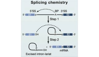
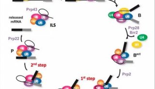
真核生物pre-mRNA剪接由超分子复合物剪接体(spliceosome)完成。完整的剪接过程主要分为八种不同的状态,预催化剪接体的前体(pre-B),预催化剪接体(B),活化复合物(Bact),催化活化复合物(B*),催化步骤I复合物 (C),催化步骤II活化复合物(C*),催化后剪接体(P)和内含子套索剪接体(ILS)。
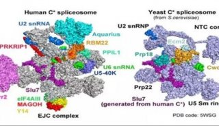
自2015年第一个近原子分辨率、来自S. pombe的剪接体结构通过Cryo-EM技术被解析以来,已经陆续有多个不同状态的剪接体被解析。其中有13个分辨率在3.3-5.8 Å之间的酵母剪接体和11个人源剪接体已经被解析,这些剪接体分布在催化过程中的七种状态中。在此之前,只剩下B*复合物结构尚未被解析。
2019年3月14日,Cell 杂志在线online了来自施一公课题组(万蕊雪和白蕊为共同第一作者)关于酵母剪接体的研究长文,题为Structures of the Catalytically Activated Yeast Spliceosome Reveal the Mechanism of Branching。这篇文章报道了2.9–3.8 Å酵母剪接体的B*复合物Cryo-EM结构,完成了剪接催化过程中几个主要步骤的最后一块拼图。

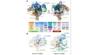
在这项研究中,施一公课题组组装了来自酿酒酵母的含有两种不同的pre-mRNA的B*复合物,其中在ACT1 pre-mRNA上组装的B*复合物平均分辨率2.9 Å;在UBC4 pre-mRNA上组装的B*复合物平均分辨率3.2 Å。对于这两种不同的B*复合物中的每一种,又观察到了两种不同的构象状态(图1 A和B)。不同pre-mRNA上的这些结构揭示了在主要功能状态下剪接体的底物特异性构象,为揭示对branching反应的机理提供了重要参考。

图1. 酵母中B*复合物结构
原文链接:
https://doi.org/10.1016/j.cell.2019.02.006
附施一公实验室有关剪接体的论文列表:
1.Yan, C. et al. Structure of a yeast spliceosome at 3.6-angstrom resolution. Science 349, 1182-1191 (2015).
2.Hang, J., Wan, R., Yan, C. & Shi, Y. Structural basis of pre-mRNA splicing. Science 349, 1191-1198 (2015).
3. Wan, R. et al. The 3.8 Å structure of the U4/U6. U5 tri-snRNP: Insights into spliceosome assembly and catalysis. Science, aad6466 (2016).
4. Yan, C., Wan, R., Bai, R., Huang, G. & Shi, Y. Structure of a yeast activated spliceosome at 3.5 Å resolution. Science 353, 904-911 (2016).
5. Wan, R., Yan, C., Bai, R., Huang, G. & Shi, Y. Structure of a yeast catalytic step I spliceosome at 3.4 Å resolution. Science 353, 895-904 (2016).
6.Yan, C., Wan, R., Bai, R., Huang, G. & Shi, Y. Structure of a yeast step II catalytically activated spliceosome. Science 355, 149-155 (2017).
7.Zhang, X. et al. An atomic structure of the human spliceosome. Cell 169, 918-929. e914 (2017).
8.Wan, R., Yan, C., Bai, R., Lei, J. & Shi, Y. Structure of an intron lariat spliceosome from Saccharomyces cerevisiae. Cell 171, 120-132. e112 (2017).
9. Bai, R., Yan, C., Wan, R., Lei, J. & Shi, Y. Structure of the post-catalytic spliceosome from Saccharomyces cerevisiae. Cell 171, 1589-1598. e1588 (2017).
10. Shi, Y. Mechanistic insights into precursor messenger RNA splicing by the spliceosome. Nature Reviews Molecular Cell Biology 18, 655 (2017).
11.Shi, Y. The spliceosome: a protein-directed metalloribozyme. Journal of molecular biology 429, 2640-2653 (2017).
12. Zhan, X., Yan, C., Zhang, X., Lei, J. & Shi, Y. Structure of a human catalytic step I spliceosome. Science 359, 537-545 (2018).
13. Bai, R., Wan, R., Yan, C., Lei, J. & Shi, Y. Structures of the fully assembled Saccharomyces cerevisiae spliceosome before activation. Science, eaau0325 (2018).
14. Zhang, X. et al. Structures of the human spliceosomes before and after release of the ligated exon. Cell research, 1 (2019).
15. Yan, C., Wan, R. & Shi, Y. Molecular mechanisms of pre-mRNA splicing through structural biology of the spliceosome. Cold Spring Harbor perspectives in biology 11, a032409 (2019).
16. Wan, R., Bai, R., Yan, C., Lei, J. & Shi, Y. Structures of the Catalytically Activated Yeast Spliceosome Reveal the Mechanism of Branching. Cell (2019).
-
焦点事件

-
焦点事件
-
焦点事件

-
焦点事件

-
焦点事件