Application Note: Qdot® Nanocrystal Conjugates in Flow Cytometry
实验概要
Researchers today are trying to maximize the information that they get out of flow cytometry experiments by looking at more parameters in a single sample. Qdot® nanocrystals provide a powerful way to multiply fluorophore selection using commonly available excitation sources. Invitrogen currently offers a growing selection of antibody conjugates using Qdot® 565, Qdot® 605, Qdot® 655, Qdot® 700, and Qdot® 800 nanocrystals.
实验原理
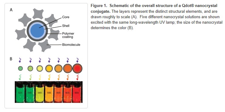
Qdot® nanocrystal conjugates are being used increasingly in multispectral flow cytometry [1–5]. These nanocrystal conjugates allow the addition of 1–6 colors excited by a UV or violet laser, and provide the opportunity to replace problematic fluorophores excited by lasers in the blue to red region. Qdot® nanocrystals provide the additional advantages of brightness and photostability. Qdot® nanocrystals are nanometer-scale semiconductor particles comprising a core, shell, and coating (Figure 1a). The core is composed of a few hundred to a few thousand atoms of a semiconductor material, often cadmium mixed with sulfur, selenium, or tellurium. The core is coated with a semiconductor shell, typically ZnS, to improve the optical properties of the material. The core and shell are encased in an amphiphilic polymer coating to provide a water soluble surface, which is covalently modified with a functionalized polyethylene glycol (PEG) outer coating. The PEG surface has been shown to reduce nonspecific binding in flow cytometry, thereby improving signal-to-noise ratios and providing clearer resolution of cell populations. Finally, antibodies are conjugated to the PEG layer using sulfhydryl/maleimide chemistry.

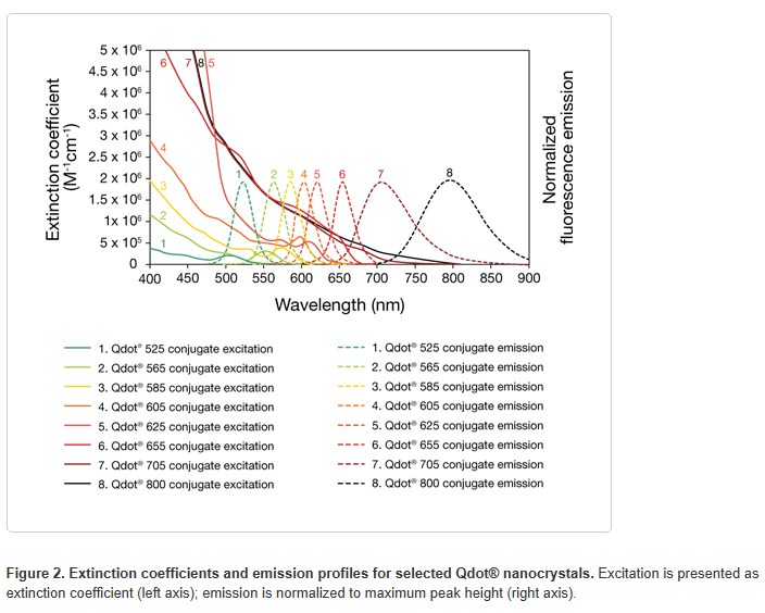
The fluorescence properties of Qdot® nanocrystals are different than those of typical dye molecules. The color of light that the Qdot® nanocrystal emits is strongly dependent on the particle size, creating a common platform of labels that ranges from green to red, all manufactured from the same underlying semiconductor material (Figure 1b). Conventional fluorophores such as fluorescein and R-phycoerythrin (R-PE) have excitation and emission spectra with relatively small Stokes shifts, which means that the emission maximum for the fluorophore is generally within 20–50 nm of the excitation maximum. Qdot® nanocrystals have symmetrical and relatively narrow emission peaks that can be 150 to 400 nm above their excitation wavelengths (Figure 2). By using nanocrystals with emission peaks that are separated by at least 40 nm (Qdot® 565, Qdot® 605, Qdot® 655, Qdot® 700, and Qdot® 800 nanocrystals), conjugates can be made that have minimal spectral overlap.

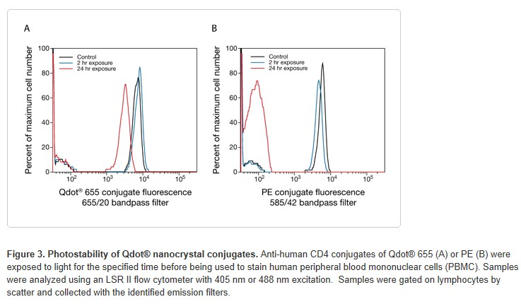
Unlike conventional dyes, Qdot® conjugates remain fluorescent under constant illumination while conventional dyes photobleach to different extents (Figure 3). The high fluorescence stability of a Qdot® nanocrystal results in better reagent stability, and an increase in the fluorescence stability of stained samples facilitates reanalysis after sorting.

主要试剂
1. Qdot® nanocrystal antibody conjugates
2. Conventional fluorophore-antibody conjugates
3. Sample: cells from blood, tissue culture, or singulated tissue
4. Sample preparation reagents:
1) Cal-Lyse™ solution (Invitrogen), or equivalent red cell lysis reagent
2) IC Fixation Buffer (Invitrogen), fixative from FIX & PERM® (Invitrogen),
% buffered formaldehyde, or equivalent cell fixation reagent
3) IC Permeabilization Buffer (Invitrogen), permeabilizer from
FIX & PERM® (Invitrogen), or equivalent cell permeabilizing reagent
5. Staining buffer: phosphate-buffered saline with 1% BSA (PBS/1% BSA), or equivalent

实验步骤
1. Collect whole blood or prepare a cell suspension in a suitable staining buffer such as PBS/1% BSA.
2. Add 100 μL cell suspension (at 1 x 10⁶ cells/mL) or 100 μL whole blood to a 12 x 75 mm flow cytometry tube.
3. Add the antibody conjugates together at optimal concentrations,including Qdot® antibody conjugate at optimal concentration, and incubate either at room temperature for 15 minutes or on ice for 30 minutes. Qdot® antibody conjugates may be added separately or mixed with other antibody conjugates.
4. Wash the cells twice with staining buffer by centrifugation and resuspend in 100 μL of staining buffer for analysis.
注意事项
1. Like any antibody conjugate, a Qdot® nanocrystal conjugate should be titered to determine its optimal staining concentration.
2. Qdot® conjugates should be diluted in staining buffer, and diluted solutions should be used within the day of dilution. Any leftover diluted stock should be discarded.
3. Antibody conjugates may be added separately or mixed and added as a cocktail. Cocktails containing Qdot® nanocrystal conjugates should be used within the day of dilution.
4. The sample may be fixed with IC Fixation Buffer for 15 minutes at room temperature. It is best to resuspend the sample in staining buffer after fixation, as long-term exposure to aldehyde-based fixatives can alter fluorochrome and cell scatter properties.
附 件 (共4个附件,占253KB)
![]()
4.jpg
59KB 查看![]()
2.jpg
83KB 查看![]()
3.jpg
63KB 查看![]()
1.jpg
48KB 查看


