活细胞成像在中枢神经系统(CNS)疾病和紊乱研...(三)

| Huvec细胞: Huvec细胞(固定),用SiR-actin染色,共聚焦显微镜成像。 | |
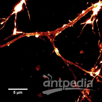 |
大鼠海马神经元: 用SiR-actin染色培养大鼠海马神经元的STED图像。肌动蛋白环(条纹)周期性为180nm。 |
 |
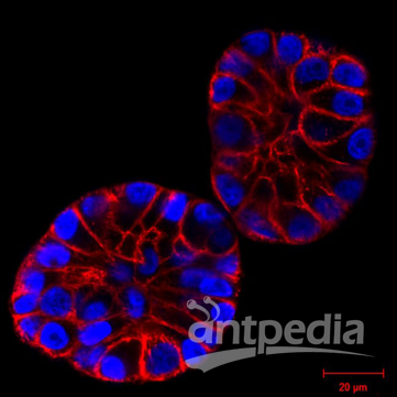
MCF10A Cells (3D培养) 用SiR -actin(红色)染色的MCF10A细胞在基质胶中表达H2B-GFP(蓝色)的。 LSM 倒置显微镜观测图 |
 |
MCF10A Cells (3D培养) 用SiR -actin(红色)染色的mcf10a细胞在基质胶中表达H2B-GFP(蓝色)的。 LSM 倒置显微镜观测图 |
 |
金鱼视网膜双极细胞: 用SiR-tubulin染色的单个分离金鱼视网膜双极细胞的三维投影。颜色光谱代表深度。可见微管从树突的顶端(顶部)伸入轴突,向下伸入巨大的突触末端(底部)。 |
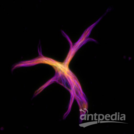 |
金鱼星形胶质细胞: 用SiR-tubulin染色的单个分离的金鱼星形胶质细胞的三维投影。 |
G-LISA 活化检测试剂盒:
| 产品名称 | 货号 |
| RhoA G-LISA 活化检测生化试剂盒(比色法) | (Cat. # BK124) |
| RhoA G-LISA 活化检测生化试剂盒(荧光法) | (Cat. # BK121) |
Actin生化试剂盒:
| 产品名称 | 货号 |
| 肌动蛋白结合蛋白 Spin-Down生化检测试剂盒(兔骨骼肌actin) | (Cat. # BK001) |
| 肌动蛋白结合蛋白 Spin-Down生化检测试剂盒(人血小板actin) | (Cat. # BK013) |
| Actin聚合生化检测试剂盒(荧光法:兔骨骼肌actin) | (Cat. # BK003) |
| G-Actin/F-actin In Vivo生化检测试剂盒 | (Cat. # BK037) |
Tubulin生化试剂盒:
| 产品名称 | 货号 |
| Tubulin聚合生化检测试剂盒(比色法) | (Cat. # BK006P) |
| Tubulin聚合生化检测试剂盒(荧光法) | (Cat. # BK011P) |
| 微管结合蛋白 Spin-Down生化检测试剂盒 | (Cat. # BK029) |
| 微管/Tubulin In Vivo生化检测试剂盒 | (Cat. # BK038) |
iRegene的人源干细胞株及配套培养试剂盒:
| 产品名称 | 货号 |
| NouvNe u™人源神经干细胞(hNSC)细胞株 | (RJC02006) |
| NouvNeu!" hNSC 神经干细胞培养试剂盒 | (RJM02000) |
| NouvNeu™hNeuron神经元定向分化细胞培养试剂盒 |
(RJM03000) |
StressMarq的活性α突触蛋白和Tau蛋白,便于快速构建神经疾病动物模型:
| 产品名称 | 活性 | 应用类型 | 货号 | 适用物种 |
| 重组人α-突触核蛋白单体(对照) 重组人α-突触核蛋白聚合体PFFs(对照) | Inactive | WB、SDS-PAGE、in vivo/vitro | SPR-316B SPR-317B | |
| 重组人α-突触核蛋白单体 重组人α-突触核蛋白聚合体PFFs | Active | WB、SDS-PAGE、in vivo/vitro | SPR-321B SPR-322B | |
| 重组小鼠α-突触核蛋白单体 重组小鼠α-突触核蛋白聚合体PFFs | Active | WB、SDS-PAGE、in vivo/vitro | SPR-323B SPR-324B | |
| 小鼠抗小鼠α-突触核蛋白单抗 小鼠抗小鼠α-突触核蛋白单抗 小鼠抗小鼠α-突触核蛋白单抗 | WB、ICC/IF WB、IHC、ICC/IF WB、IHC、ICC/IF | SMC-531DSMC-532DSMC-533D | 人、小鼠、大鼠 人、小鼠、大鼠 人、小鼠、大鼠 | |
| 小鼠抗人α-突触核蛋白单抗 兔抗人α-突触核蛋白多抗 | WB、ICC/IF WB | SMC-530DSPC-800D | 人、小鼠、大鼠 | |
| α-突触核蛋白 (磷酸化Ser129) 抗体 α-突触核蛋白 (磷酸化Tyr136) 抗体 | WB、ICC/IF WB | SPC-742DSPC-1435D | 人、小鼠 人、小鼠、大鼠 | |
| 重组Tau 2N4R P301S蛋白单体 重组Tau 2N4R P301S蛋白PFFs | Active | WB、SDS-PAGE、in vivo/vitro | SPR-327B SPR-329B | |
| 重组Tau K18/P301L蛋白单体 重组Tau K18/P301L蛋白PFFs | Active | WB、SDS-PAGE、in vivo/vitro | SPR-328B SPR-330B |
参考文献:
Neurological disease models made clear. Med.(Editorial, published September 2015). 21, 964.
Schlachetzki J.C. et al. 2013. Studying neurodegenerative diseases in culture models. Bras. Psiquiatr.35, S92-100.
Hughes P. et al. 2018. The costs of using unauthenticated, over-passaged cell lines: how much more data do we need? Biotechniques. 43, 575, 577-578, 581-582
Verstraelen P. et al. 2018. Image-based profiling of synaptic connectivity in primary neuronal cell culture. Neurosci. 12, 389.
Hoover B.R. et al. 2010. Tau mislocalization to dendritic spines mediates synaptic dysfunction independently of neurodegeneration. Neuron. 68, 1067-1081
Spires-Jones T.L. and Hyman B.T. 2014. The intersection of amyloid beta and tau at synapses in Alzheimer’s disease. Neuron. 82, 756-771.
Bayer T.A. and Wirths O. 2010. Intracellular accumulation of amyloid-beta – a predictor for synaptic dysfunction and neuron loss in Alzheimer’s disease. Front. Aging Neurosci. 2, 8
Unternaehrer J.J. and Daley G.Q. 2011. Induced pluripotent stem cells for modelling human diseases. Trans. R. Soc. B.366, 2274-2285.
Munoz S.S. et al. 2018. The serine protease HtrA1 contributes to the formation of an extracellular 25-kDa apolipoprotein E fragment that stimulates neuritogenesis. Biol. Chem. 293, 4071-4084
Jin M. et al. 2018. An in vitro paradigm to assess potential anti-Aβ antibodies for Alzheimer’s disease. Commun.9, 2676.
Hong W. et al. 2018. Diffusible, highly bioactive oligomers represent a critical minority of soluble Aβ in Alzheimer’s disease brain. Acta Neuropathol.136, 10-40
Di Primio C. et al. 2017. The distance between N and C termini of tau and of FTDP-17 mutants is modulated by microtubule interactions in living cells. Neurosci.10, 210.
White M.D. et al. 2018. In vivo imaging of single mammalian cells in development and disease. Trends Mol. Med.24, 278-293.
Wrasidlo W. et al. 2016. A de novo compound targeting alpha-synuclein improves deficits in models of Parkinson’s disease. 139, 3217-3236.
Falkner S. et al. 2016. Transplanted embryonic neurons integrate into adult neocortical circuits. Nature. 539, 248-253.
Peron S. et al. 2017. A delay between motor cortex lesions and neuronal transplantation enhances graft integration and improves repair and recovery. Neurosci.37, 1820-1834.
Wang C. et al. 2016. Infiltrating cells from host brain restore the microglial population in grafted cortical tissue. Rep.6, 33080.
Guo L. et al. 2015. Dynamic rewiring of neural circuits in the motor cortex in mouse models of Parkinson’s disease. Neurosci.18, 1299-1309.
Shin H.Y. et al. 2018. Using automated live cell imaging to reveal early changes during human motor neuron degeneration. eNeuro. doi: 10.1523/ENEURO.0001-18.2018.
-
焦点事件