


Embryonic limb bud culture in media
Early in embryonic development, the region of the chick embryo which is determined to form a limb first differentiates from the rest of the embryo1. The next step in the formation of the actual limb (wing or leg) is the development of the limb region into a specific set of functional tissues through the interactions of several proteins, or morphogenesis. Morphogenesis of the vertebrate limb occurs similarly in all tetrapods. This similarity is marked by growth in three dimensions which are along the proximal-distal, anterior-posterior, and dorsal-ventral axes.
By the sixth day, the limb bud possesses all the necessary signals to continue growth of the limb in culture and is visible as a process on the embryo with distinct axes. As development continues, the basic limb structure arises out of the limb bud as skin, cartilage, and skeletal muscle. As the embryo matures, the cartilage will turn into bone, thus getting closer to fulfilling the fate which began in the early embryo. In addition to differentiating into basic structure, the limbs differentiate from each other. This differentiation is determined by the interaction of several chemicals and growth factors. As a result, a wing is not the same as the leg. The consequence of normal limb development in the chick embryo are the legs and wings necessary for the organism's survival.
Two methods of limb bud culture exist. Chorioallantioc membrane (CAM) grafting is a culture method commonly used in the past. In this type of experiment, the severed limb bud is grafted into a host egg on a Y- shaped junction of blood vessels. The limb grows on nutrients supplied by the host egg. A more recent application of limb bud uses no host but a growth medium in vitro. The culture medium contains elements essential for continued growth and antibiotics to protect against infection. Although CAM grafts have been used in the past, an obvious advantage of an in vitro method is that manipulative agents, such as growth factors and hormones, can be added to the culture medium.
The primary purpose of this experiment is to observe limb development by studying the presence of cartilage in cultured limbs. The secondary purpose is to investigate whether or not in vitroculture can support limb development. The control will be limbs that develop in ovo.
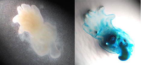
Major Observations
Limbs developed in vitroand cartilage was observed using Alcian green staining. In some cases, development had progressed to the point at which cartilage began changing into bone, and, thus, was not seen in the staining. Compared to control limbs, limbs grown in media were not as large or organized as well.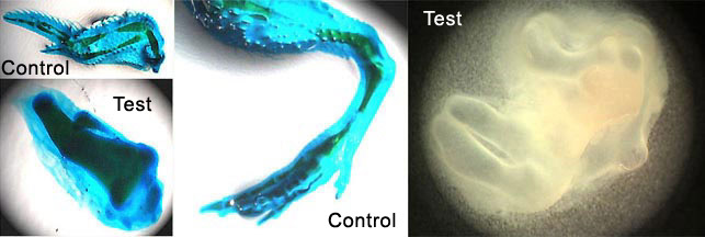
Detailed Laboratory Procedures2 Week 1: Preparation and culture of the limb bud.
1. In 6 well plates, place tissue culture filter inserts over approximately 1.8ml of culture media. | |||||||||
Week 2: Staining for limb cartilage. | |||||||||
1. Fix limbs in 5% TCA for 1 hour at room temperature. Culture media:
| |||||||||
