凋亡细胞核DNA片段检测方法进展(二)
2.3
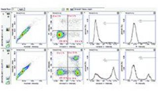
凋亡细胞的TUNEL和ISNT鉴定的流式细胞仪分析[16]对于培养的细胞,可以将TUNEL或ISNT鉴定同流式细胞仪结合起来分析其发生凋亡的情况。待检细胞与含有TdT或DNA聚合酶I或Klenow片段及生物素标记的dUTP反应液共孵育一段时间后,加入荧光素(常用FITC)标记的链霉抗生物素蛋白(streptavidin),使之特异性地与挂在DNA断链上的生物素结合,然后在流式细胞仪上分析荧光强度,可结合其他的荧光染色方法(如PI染色和荧光免疫表型鉴定)行多参数的流式细胞仪分析,使之有很广的应用价值。该方法的主要优点有:可根据荧光强度对凋亡细胞作出定量分析;可以区别凋亡和坏死;结合表型鉴定,确定不同来源的细胞群中某一或几种细胞亚群的凋亡;其检测与凋亡相关的DNA降解片段的特异性和敏感性比琼脂糖凝胶电泳高得多,能检测出少于(2~5)×103个细胞的DNA断链。
3 细胞凋亡的ELISA检测[17]
细胞凋亡ELISA检测就是基于酶联免疫吸附夹心法测定原理,用单克隆抗DNA抗体和抗组蛋白抗体直接检测DNA和组蛋白,可特异性定量检测细胞溶解物细胞浆成分中的单和寡核苷酸小体。其优点有:无需使用放射性同位素;可对细胞凋亡进行定量检测;抗组蛋白抗体无物种的特异性,可用于各种物种的细胞凋亡的检测;高敏感性,较少的细胞就可获得结果。
4 结语
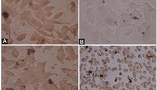
琼脂糖凝胶电泳简单、方便,出现“梯状”带虽能证实凋亡,但只能定性不能定量,不是随时电泳都能得到“梯状”带,因而敏感性不高,且这种方法一般只用于培养的细胞,而组织的异源性不能保证该方法的特异性;其他一些电泳方法如反转电场凝胶电泳和彗星鉴定也有其自身的应用条件,针对性较强。原位末端标记法的出现,是细胞凋亡方法学上的一大进步,特别是经众多学者的应用和完善,其特异性尤其是敏感性方面有了很大的提高,广泛应用于各种组织、细胞培养的细胞凋亡的研究,特别是结合抗原免疫表型鉴定和流式细胞仪分析,可以区分不同来源的组织或细胞中的某种或几种细胞亚群的凋亡,并进行定量研究。正如上面所说,该方法的特异性有一定的限度,就其技术本身而言,并不能区别凋亡、坏死和自溶性死亡,必须结合凋亡细胞的形态学特征加以判断。因此,在细胞凋亡的研究中,多种方法的结合是必要的,也是必须的。
作者简介:谭晓华,男,1962年出生,博士,主治医师作者单位:姜泊 张亚历 周殿元 (第一军医大学南方医院消化内科研究所,广州,510515)
谭晓华 (北京军区总医院肝病研究所)
参考文献
1 Oberhammer F,Wilson JW,Dive C et al.Apoptotic death in epithelial
cells: cleavage of DNA to 300 and/or 50 kb fragments prior to or in the
absence of internucleosomal fragmentation.EMBO J,1993,12:3679
2 Bicknell GR,Cohen GM.Cleavage of DNA to large kilobase pair fragments
occurs in some forms of necrosis as well as apoptosis.Biochem Biophys
Res commun,1995,207:40
3 Cohen GM,Sun XM,Snowden RT et al.Key morphological features of
apoptosis may occur in the absence of internucleosomal DNA
fragmentation.Biochem j,1992,286:331
4张亚历,姜泊,周殿元.程序化细胞死亡常用研究方法.见:姜泊等主编.分子生物学常用实验方法.北京:人民军医出版社,1996.170~175
5 谭晓华,张亚历,姜泊等.凋亡细胞中小片段DNA的简单快速提取方法.第一军医大学学报,1996,16(3):227
6 Sestili P,Cattabeni F,Cantoni O.Direct excision of 50 kb pair DNA
fragments from megabase- sized fragments produced during apoptotic
cleavage of genomic DNA.FEBS Lett,1996,396:337
7 Olive PL,Frazer G,Banath JP.Radiation-induced apoptosis measured in
tK6 human B lymphoblast cells using the comet assay.Radiat
Res,1993,136:130
8 Huang P,Plunkett W.A quantitative assay for fragmented DNA in apoptotic cells.Anal Biochem,1992,207:163
9 Negoescu A,Lorimier P,Labat Moleur F et al.In situ apoptotic cell
labeling by the TUNEL method: improvement and evaluation on cell
preparations.J histochem Cytochem,1996,44:959
10 Migheli A,Cavalla P,Marino S et al.A study of apoptosis in normal and
pathologic nervous tissue after in situ end-labeling of DNA strand
breaks.J neuropathol Exp Neurol,1994,53:606
11 Grasl KB,Ruttkay NB,Koudelka H et al.In situ detection of fragmented
dNA (TUNEL assay) fails to discriminate among apoptosis,necrosis,and
autolytic cell death: a cautionary note.Hepatology,1995,21:1465
12 Ansari B,Coates PJ,Greenstein BD et al.In situ end-labelling detects
dNA strand breaks in apoptosis and other physiological and pathological
states.J pathol,1993,170:1
13 Wijsman JH,Jonker RR,Keijzer R et al.A new method to detect apoptosis
in paraffin sections: in situ end-labeling of fragmented DNA.J
histochem Cytochem,1993,41:7
14 Gold R,Schmied M,Rothe G et al.Detection of DNA fragmentation in
apoptosis:application of in situ nick translation to cell culture
systems and tissue sections.J Histochem Cytochem,1993,41:1023
15 谭晓华,周殿元,姜泊等.两种原位DNA断链标记技术检测组织切片和培养细胞中的凋亡细胞.中华医学检验杂志,1998,21(9):278
16 Dolzhanskiy A,Basch RS.Flow cytometric determination of apoptosis in
heterogeneous cell populations.J Immunol Methods,1995,180:131
17 Salgame P,Varadhachary AS,Primiano LL et al.An ELISA for detection of apoptosis.Nucleic Acids Res,1997,25:680