Em observations of microsomes
Em observations of microsomes
LEVEL I

Figure 7.3 Isolated microsomes

MATERIALS
1% Glutaraldehye (GTA)
1% Osmium tetroxide
Epoxy or vinyl resin for TEM
TEM photomicrograph of liver cells
Transmission electron microscope
PROCEDURE
Place a mm
 piece of the final pellet from Exercise 7.1 into a small vial containing 1% GTA. Fix the pellet for 2 hours.
Rinse the pellet with water and place in three changes of water for 30 minutes each, to remove any residual GTA.
Post fix the lysosome pellet in 1% osmium tetroxide for 1 hour. Wash thoroughly with 3 changes of water (30 minutes each).
Dehydrate the tissue by passing through a series of graded alcohols or acetone, and embed in plastic blocks.
Section the plastic blocks and place on coated grid. Double stain with uranyl acetate and lead nitrate and examine with a transmission electron microscope.
Compare the view of the compacted lysosome pellet to the structure and distribution of microsomes in an intact hepatocyte (liver cell). Use figures 7.3 and 7.4 for reference.
-
项目成果
-
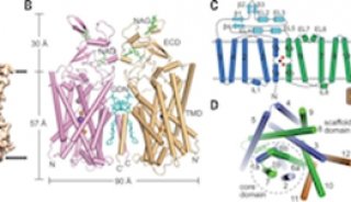
项目成果
-
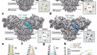
项目成果
-
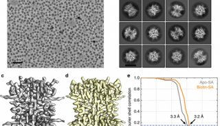
产品技术