ASTM E1588-10e1
扫描电子显微术/能量散射X射线光谱法射击残留物分析的标准指南
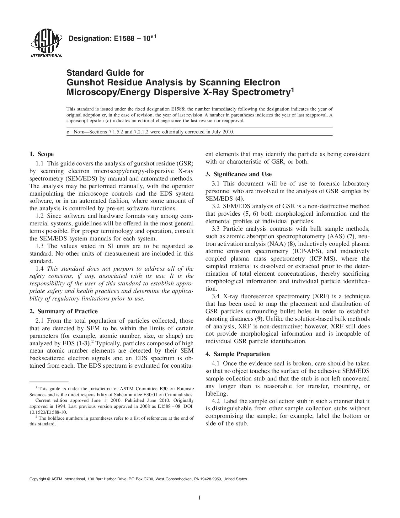
Standard Guide for Gunshot Residue Analysis by Scanning Electron Microscopy/ Energy Dispersive X-ray Spectrometry
- 标准号
- ASTM E1588-10e1
- 发布
- 2010年
- 发布单位
- 美国材料与试验协会
- 替代标准
- ASTM E1588-16
- 当前最新
- ASTM E1588-20
- 适用范围
This document will be of use to forensic laboratory personnel who are involved in the analysis of GSR samples by SEM/EDS (4).
SEM/EDS analysis of GSR is a non-destructive method that provides (5, 6) both morphological information and the elemental profiles of individual particles.
Particle analysis contrasts with bulk sample methods, such as atomic absorption spectrophotometry (AAS) (7), neutron activation analysis (NAA) (8), inductively coupled plasma atomic emission spectrometry (ICP-AES), and inductively coupled plasma mass spectrometry (ICP-MS), where the sampled material is dissolved or extracted prior to the determination of total element concentrations, thereby sacrificing morphological information and individual particle identification.
X-ray fluorescence spectrometry (XRF) is a technique that has been used to map the placement and distribution of GSR particles surrounding bullet holes in order to establish shooting distances (9). Unlike the solution-based bulk methods of analysis, XRF is non-destructive; however, XRF still does not provide morphological information and is incapable of individual GSR particle identification.
1.1 This guide covers the analysis of gunshot residue (GSR) by scanning electron microscopy/energy-dispersive X-ray spectrometry (SEM/EDS) by manual and automated methods. The analysis may be performed manually, with the operator manipulating the microscope controls and the EDS system software, or in an automated fashion, where some amount of the analysis is controlled by pre-set software functions.
1.2 Since software and hardware formats vary among commercial systems, guidelines will be offered in the most general terms possible. For proper terminology and operation, consult the SEM/EDS system manuals for each system.
1.3 The values stated in SI units are to be regarded as standard. No other units of measurement are included in this standard.
1.4 This standard does not purport to address all of the safety concerns, if any, associated with its use. It is the responsibility of the user of this standard to establish appropriate safety and health practices and determine the applicability of regulatory limitations prior to use.
ASTM E1588-10e1相似标准
推荐
常见仪器原理及其所提供信息。。。
;扫描电子显微术SEM☆分析原理:用电子技术检测高能电子束与样品作用时产生二次电子、背散射电子、吸收电子、X射线等并放大成像 ☆谱图的表示方法:背散射象、二次电子象、吸收电流象、元素的线分布和面分布等 ☆提供的信息:断口形貌、表面显微结构、薄膜内部的显微结构、微区元素分析与定量元素分析等 原子吸收AAS☆原理:通过原子化器将待测试样原子化,待测原子吸收待测元素空心阴极灯的光,从而使用检测器检测到的能量变低...
实验员必备的材料成分分析技术大全
:晶体形貌、分子量分布、微孔尺寸分布、多相结构和晶格与缺陷等扫描电子显微术 SEM分析原理:用电子技术检测高能电子束与样品作用时产生二次电子、背散射电子、吸收电子、X射线等并放大成象谱图的表示方法:背散射象、二次电子象、吸收电流象、元素的线分布和面分布等提供的信息:断口形貌、表面显微结构、薄膜内部的显微结构、微区元素分析与定量元素分析等原子吸收 AAS原理:通过原子化器将待测试样原子化,待测原子吸收待测元素空心阴极灯的光...
材料成分分析技术大全,实验员快收藏!
:晶体形貌、分子量分布、微孔尺寸分布、多相结构和晶格与缺陷等扫描电子显微术 SEM分析原理:用电子技术检测高能电子束与样品作用时产生二次电子、背散射电子、吸收电子、X射线等并放大成象谱图的表示方法:背散射象、二次电子象、吸收电流象、元素的线分布和面分布等提供的信息:断口形貌、表面显微结构、薄膜内部的显微结构、微区元素分析与定量元素分析等原子吸收 AAS原理:通过原子化器将待测试样原子化,待测原子吸收待测元素空心阴极灯的光...
材料成分分析技术大全
:晶体形貌、分子量分布、微孔尺寸分布、多相结构和晶格与缺陷等扫描电子显微术 SEM分析原理:用电子技术检测高能电子束与样品作用时产生二次电子、背散射电子、吸收电子、X射线等并放大成象谱图的表示方法:背散射象、二次电子象、吸收电流象、元素的线分布和面分布等提供的信息:断口形貌、表面显微结构、薄膜内部的显微结构、微区元素分析与定量元素分析等原子吸收 AAS原理:通过原子化器将待测试样原子化,待测原子吸收待测元素空心阴极灯的光...

