In VitroCulture of Chicken Heart
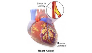
The chicken is a classic organism used to illustrate the principles of basic embryology. One of the developmental systems which has been examined in great detail is the circulatory system. In the developing embryo, the circulatory system is the first functional unit and the heart is the first functional organ. The heart of the chicken embryo develops from the fusion of paired precardiac mesodermal tubes, forming a straight anterior to posterior ventricular tube. After fusion is complete, the heart tube has four distinct regions:bulbus cordis, ventricle, atrium, and sinus venosus. Pulsations in the heart starts while the paired primordial cells fuse. The sinus venosus is the pacemaker of these initial contractions (Gilbert 1997).
After approximately 33 hours the heart tube bends to form an "S" shape structure with a single atrium and a single ventricle. By 2 days the heart has folded upon itself forming a single loop. This moves the sinus venosus and atrium to a position anterior and dorsal to the ventricle and the bulbus cordis. In 3 day-chick embryos, the atrium has begun to expand to the left in preparation of the division into the right and left atria. Although the heart still has two chambers at this time, communication between the sinus venosus and the atrium occurs through the right side of the atrium. Times of development may vary. Eventually, when the atrium and ventricle divide to develop a typical four-chambered heart, the sinus venosus will be incorporated into the right atrium. The bulbus cordis will eventually give rise to the aorta.
In this experiment, we will remove hearts from 2-day and 3-day chick embryos (see below) and maintain them under specific conditions which allow for their development. By this time the two chambered heart should be visible as well as the blood flow entering the lower chamber and being pumped out through the aorta. The goals of this lab are:to identify the anatomy of the developing chick heart, determine the direction of blood flow through the developing heart, and to observe how the heart develops over time.
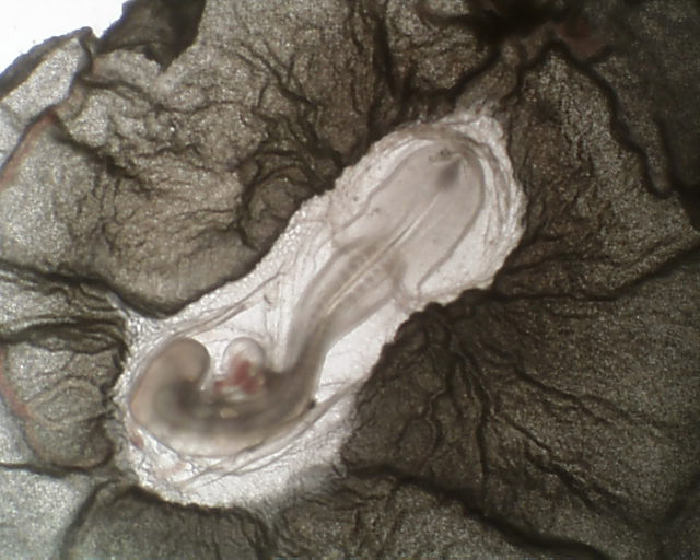
3 day chick embryo heart donor
Major Observations
All the 2 day hearts stopped beating after the first day of removal. However, the 3 day hearts continued to develop and beat in culture for approximately 2 days after removal. Therefore, Looping and thickening of the atrium can be observed as well as the direction of blood flow (see movie below).
Solutions
Howard Ringer's solution
7.2 gNaCl
0.17 gCaCl2(anhyd.) or .23 g CaCl2.2H2O
0.37 gKCl
add dH2O to 1 liter
-
焦点事件