


Cajal Body Isolation Protocol
Buffers and solutions
(All solutions are supplemented with Complete Protease inhibitor tablet (Roche, Cat no: 1-873-580) at the final concentration of 1 tablet/50ml):
S1 solution: 0.25M sucrose, 10 mM MgCl2
S2 solution: 0.35M sucrose, 0.5 mM MgCl2
S3 solution: 0.5M sucrose, 25 mM Tris-HCl pH 9.0
SP1 buffer: 1M sucrose, 34.2% Percoll, 22.2 mM Tris-HCl pH 7.4, 1.11 mM MgCl2
SP2 buffer: 20% Percoll, 10mM Tris-HCl pH 7.4, 1% Triton X100, 0.5 mg/ml heparin
HT buffer: 10mM Tris-HCl pH7.4, 1% Triton X100, 0.5 mg/ml heparin
(See “Notes” below on making sucrose stock solution)
Procedure
(All procedures should be performed either on ice or at 4°C)
|

A. SONICATION
1. Prepare the starting material of 2.5x10⁹ HeLa nuclei. Suitable HeLa nuclei can be purchased from 4C Biotech (Seneffe, Belgium; Cat no: CC-01-30-25). Add S1 solution to the thawed nuclei to a final volume of 30ml. Mix thoroughly but avoid vigorous vortexing.
2. Divide the diluted nuclei into 2x15ml portions. Overlay each portion onto 15ml of S2 solution in a 50 ml Falcon tube. Make sure the interface of the two layers is sharp (Figure 1).
 |
Figure 1: Steps 2-3 of the procedure. Note the clear boundary between the S1 and S2 layers before centrifugation.3. Centrifuge at 1430g (2500 rpm, Beckman GS-6 centrifuge, GH-3.8 rotor) for 5min at 4°C. |
4. Carefully decant the supernatant. The pellets should be solid and firm (Figure 1). Resuspend each pellet with S2 solution so that the final volume is 30ml. The pellets should resuspend easily. A pellet that cannot be resuspended completely indicates the presence of lysed nuclei and should be discarded.
5. Divide the resuspended nuclei into 10x3ml portions, each in a 15ml Falcon tube. In our experience, sonication is more effective with this sample size.
6. Sonicate each aliquot using a tuned sonicator (Misonix XL 2020) for 3x6 sec, with a 6 sec intermission between each burst. See “Notes” below on sonication.
7. Examine the sonicated nuclei under a phase contrast microscope. The majority of the nuclei should have been lysed, while the nucleoli should be clearly visible. If the percentage of unlysed nuclei is too high (over 5%), sonicate again for 2-3 sec, then re-examine under the microscope. Stop at the point when most nuclei are lysed. Over sonication disrupts Cajal bodies. The conditions used to sonicate HeLa nuclei not only maintains the structure of CBs, but also that of nucleoli, PML bodies and splicing speckles. This indicates that, under suitable conditions, many nuclear domains remain intact even after the overall nuclear structure is destroyed.
B. REMOVAL OF NUCLEOLI
8. Pool the 10 aliquots back together. Measure the volume. Add 0.42X volume of 2.55M sucrose to the sonicated nuclei so that the final sucrose concentration is now 1M. Mix thoroughly but don’t vortex. Divide the mixture into two portions, approx. 20ml each, in two 50ml Falcon tubes (Figure 2).
9. Centrifuge at 3000g (3500 rpm, Beckman GS-6 centrifuge, GH-3.8 rotor) for 10 min at 4°C.
|
Figure 2: Steps 8-10 of the procedure. Note the small pellet after centrifugation. The panels on the right show the microscope images of the supernatant and pellet. Anti-coilin antibodies (5P10, green) and Pyronin Y (red) were used to label CBs and nucleoli respectively. Bar: 10µm. |

10. A small pellet should be visible in each tube (Figure 2). This pellet contains nucleoli and unlysed nuclei, while the supernatant is nucleoplasm containing CBs. Using a pipette, carefully collect the supernatant. Do not decant, as the pellets are not firm. Stop when you are about 5mm above the pellet. This is the region where the distinction between the pellet and the supernatent is blurred. This part of the supernatant should not be included in the subsequent steps.
11. To collect nucleoli for other experiments, resuspend the pellet with 3ml S2. Sonicate the mixture for 3x10sec. Proceed from step 7 of the nucleolar isolation protocol.
C. GRADIENT ONE
12. Measure the volume of the supernatant. Add 0.82X volume of SP1. For example, in a typical experiment, mix 37ml of the supernatant with 30.27ml SP1. Add 0.05X volume of 20% Triton X100 (3.36ml in the above example). Mix thoroughly but do not vortex.
13. Divide the mixture into pre-cooled SW41 ultracentrifuge tubes (Figure 3). The volume of the sample should fit 6 tubes perfectly. Balance the tubes carefully. Load the tubes in to a pre-cooled SW41 rotor. Transfer the rotor to a pre-cooled ultracentrifuge.
14. Ultracentrifuge 37,000rpm, 2 hours.
 |
Figure 3: Steps 13-15 of the procedure. Note the loose pellet (1P) after centrifugation. Immunofluorescence (panels on the right) shows that most CBs are in 1P. Bar: 10µm. |
15. A loose pellet can be seen resting on the percoll precipitate at the bottom of each tube (Figure 3). This pellet contains most of the Cajal bodies. Carefully pipette away the turbid supernatant. Do not decant. Stop when you are 5-6mm above the pellet. Keep the supernatant labeled as Fraction 1S. Use a P1000 pipette and resuspend the pellets in the remaining supernant. Transfer the resuspended pellets to a 15ml Falcon tube. This is Fraction 1P. Measure the final volume of Fraction 1P. In a typical experiment, the final volume is about 5ml. In Fraction 1P, the CBs present were mostly entangled with large pieces of chromatin, as revealed by DAPI staining. Nucleoli that had not been removed in the first step were also a significant contaminant.
D. GRADIENT TWO
16. Add 600 units of DNase1 (Sigma Cat no: D4527; 20,000 units/ml) to Fraction 1P. Mix with a rotating wheel for 1 hour at RT.
17. Add 0.05x volume of heparin (10mg/ml). The mixture should become more transparent on mixing.
18. Add 1x volume of SP2. Mix gently with a rotating wheel for 1 min.
19. Load the mixture in pre-cooled tubes suitable for SW55 rotor (Figure 4). In a typical experiment, the mixture fits 2 tubes perfectly. Ultracentrifuge, 45,000rpm, 1 hour.
|
Figure 4: Steps 19-20 of the procedure. Note the thin band (2.2) in the middle of the gradient after centrifugation. Bar: 10µm. |

20. Three bands should be visible (Figure 4). A broad white band floats on the top, a thin white band at the middle, and a pellet resting on the Percoll precipitate at the bottom. Using a P1000 pipette, very carefully unload the gradient from the top. Take the top band, and go on until you are 2-3mm above the middle band. This is Fraction 2.1. Use a new pipette tip, and take the middle band (Fraction 2.2). Finally, resuspend the pellet using the remaining part of the gradient. This is Fraction 2.3. For each 5ml gradient, typical volumes of the 3 fractions are: Fraction 2.1: 2.5ml; Fraction 2.2: 1ml; Fraction 2.3: 1.5ml. Most of the CBs are contained in Fraction 2.2 in the middle of the gradient, where the resolution is the greatest.
E. CONCENTRATION AND FINAL ENRICHMENT OF CAJAL BODIES
21. Into Fraction 2.2, add 10x volume of HT buffer. Mix well using a rotating wheel.
22. Either: Load the diluted fraction into 2xSW41 tubes. Ultracentrifuge. 12,000rpm, 15 min.
Or: Divide the mixture into 20x1.5ml Eppendorf tubes. Centrifuge in an Eppendorf centrifuge, 14,000rpm, 15min (Figure 5).
|
Figure 5: Step 22-27 of the procedure. Most CBs are found in Fraction 3S. Arrows in the inset indicate the presence of small amount of coilin-negative material in 3S. Bars: Big inset: 25µm, small insets: 10µm. |

23. Carefully remove the supernatant. Resuspend the pellet in 0.5ml HT buffer. Pool all the resuspended pellets into a single tube, and centrifuge again as above. The final pellet should be visible, but very small. Carefully remove the supernatant.
24. Resuspend the pellet with 0.5ml S3 solution. The pellet detaches from the bottom of the tube and is resuspended on pipetting up and down.
25. Spin down the resuspended pellet in an Eppendorf centrifuge, 8000rpm, 5min. A visible pellet should be seen. Carefully take the supernatant and transfer it to a new Eppendorf tube. Repeat spinning the supernatant at 8000rpm, 5min. Carefully take the supernatant and transfer it to a new Eppendorf tube.
26. Combine the pellets. Label this as Fraction 3P.
27. Dilute the supernatant with 10x volumes of 25mM Tris-HCl pH 9.0. Divide the diluted supernatant into 3x1.5ml Eppendorf tubes. Centrifuge at 14,000rpm, 15min. Carefully remove the supernatants, leaving about 0.1ml at the bottom of each tube. Pipette the remaining supernatants up and down, resuspending the small pellet. Mix the content of the 3 tubes together. Fill up the tube with 25mM Tris-HCl pH 9.0, and centrifuge again at 14,000rpm,15min. Carefully remove the supernatant, leaving about 20µl in the tube. Pipette the remaining supernatants up and down to resuspend the small pellet. Label this as Fraction 3S. This fraction contains enriched CBs.
Notes
(1) Making 2.55M sucrose stock
Here is a protocol for preparing a sucrose stock solution (Cline and Ryel,l 1971) suitable for the nucleolar isolation protocol. The resulting solution is 2.55M, or 66% by weight. Its density is 1.3224g/cm3 at 20oC, and refractive index is1.4558. The stock solution is stable indefinitely at 4oC. This procedure can be carried out at RT. There is no need to heat up the solution to help dissolving the sucrose. Heating up an incompletely dissolved sucrose solution can lead to charring of sucrose and affect the quality of the sucrose solution.
1. Weigh out 1710 g sucrose (BDH). Keep it aside in a clean container.
2. Put exactly 900ml water and a magnetic bar in a 5 litre beaker. Put the beaker on a stirrer and start stirring.
3. Add 1/3 of the sucrose into the beaker. Make sure the magnetic bar is rotating freely. Stir for 1 hour.
4. Add another 1/3 of the sucrose into the solution. Again make sure the rotation of the stir bar is not impaired. Stir for another 1 hour.
5. Add the remaining sucrose. Stir for another 1 hour, or until all the sucrose has gone into solution. The final volume should be exactly 2 litres.
(2) Sonication
We use a Misonix 2020 sonicator fitted with a microtip at power setting 5. To ensure reproducible soncation these points should be followed:
- It is necessary to tune the sonicator every time after you change the probe. Follow the manufacturer’s manual for the tuning procedure.
- Sonication produces intense and localized heat in your solution. If you are concerned about the heating, the correct way to reduce heating is to shorten the sonication time and to increase the intermission between bursts. Keeping the tube on ice or performing the sonication in the cold room is helpful, but is not the most effective way of heat control.
- If the probe is too close to the liquid surface, it produces a foam and reduces the efficiency of sonication. Make sure the probe is well submerged in the solution, about 5mm above the bottom of the tube. Do not, however, touch the bottom or the wall of the tube with the probe.
- Sonicator probe that has been used repeatedly develops pits on its end. The sonication efficiency gradually decreases as time goes on. Therefore, the sonication time recommended here can only be used as a guideline. Always monitor the outcome of sonication using a phase contrast microscope. You may need to adjust the sonication time to maintain the efficiency, especially if the probe is getting old. Change the probe when the efficiency is noticeably down.
(3) Analysis of the enriched Cajal body fraction
- To immunolabel the isolated CBs, spot about 5 µl of Fraction 3S on to a polylysine-coated slide (BDH Cat no: 406/0178/00), and air dry the spot. Rehydrate the slide in PBS for 5 min before using standard immunostaining procedure.
- To separate and analyse the proteins by SDS PAGE, either resuspend directly in Laemmli SDS sample buffer, or in your preferred buffer.


-

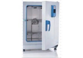
Thermo Scientific Heratherm 大容量高端型烘箱(Thermo Scientific Heratherm Large Capacity Advanced Protocol Ovens)
询底价 -
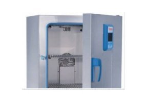
Thermo Scientific Heratherm 高端安全型烘箱(Thermo Scientific Heratherm Advanced Protocol Security Ovens)
询底价 -
Thermo Scientific Heratherm 高端型烘箱(Thermo Scientific Heratherm Advanced Protocol Ovens)
询底价 -
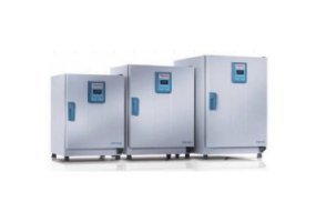
Thermo Scientific Heratherm 通用型烘箱(Thermo Scientific Heratherm General Protocol Ovens)
询底价


