荧光微球分析技术及荧光微球吞噬实验的操作流程
荧光 微球分析技术属于化学材料发展结果,可用于细胞表面抗原的检测、退行性神经病变示踪物、吞噬功能的检测、血流分析、敏感性诊断试剂等,本文介绍了荧光微球分析技术以及荧光微球吞噬实验的操作步骤。
荧光微球分析 技术简介
荧光微球分析技术是近年来化学材料科学活跃发展 的产物,各种大小(0.2~10μm)可产生荧光和色彩的人工微球应运而生,目前各种材料人工合成、多种颜色和规格的荧光微球有2大类,一种是表面不带修饰基团的微球,另一种是携带各种化学修饰基团,包括羧基化修饰、氨基修饰、巯基或醛巯基修饰等的微球。
应用
这些荧光微球正广泛应用于生命科学研究的多个领域中,如:
(1)细胞表面抗原的检测,包括CD4/CD8表面抗原的绝对计数;细胞表面低丰度表达受体的分析;骨髓移植受体内供体红细胞的检测;白色念珠菌抗原检测等。
(2)退行性神经病变示踪物,荧光微球具有无毒、与神经细胞结合时间长以及受注射部位影响极小的特点。
(3)吞噬功能的检测,0.6~2.0μm大小的荧光微球适合于这一类的研究,如分析大鼠中性粒细胞、人横纹导管细胞、小鼠腹膜巨噬细胞、人多核白细胞的吞噬功能或不同调理素调理作用对吞噬功能的影响等。
(4)血流分析,10-15 μm大小7种颜色的荧光可供研究组织中局部血流情况,如肿瘤脉管血流速率、视网膜和脉络膜循环、肺泡微管的功能直径定位等。
(5)敏感性诊断试剂,如替代一些已开展应用的微球诊断试验:胶乳凝集试验、微球捕获ELASA、双位点夹心法等,它较传统比色方法更为灵敏;另有新近诞生的流式微球分析技术(Cytometric Bead Array CBA)通过将不同荧光强度的聚苯乙烯微球包被上多种高特异性的单克隆抗体,在微球“三明治”平台上进行可溶性蛋白的检测,由此而使得1份微量标本可同时1次定量分析多种可溶性蛋白,大大节约了样本量和操作时间,灵敏度也较传统的ELISA方法更高,重复性好。
(6)质量控制,2~10 μm直径,大小均一,表面定量吸附不同荧光素的树脂颗粒可用于流式细胞仪的质控,在样品测试前调整仪器对测试信号的放大系数,以保证各次实验结果具有高度的重复性,这类微球标准品按其应用被分为以下2类:即定量荧光标准微球和准值荧光标准微球。
荧光微球吞噬实验的Protocol
---Materials
*Fluorescence labeled latex beads (1um diameter), 2.5% aqueous suspension
* 3% BSA containing 25 mM Na2HPO4, pH 6.0
* 0,3%(w/v) azide
* Culture medium containing 5% FBS
* Distilled water, PBS
* Bath sonication, 6(12)-well plates
---Cell culture and treatment
1, Inoculate plates with 7,0104 cells/cm2 per well. Incubate at 37℃,5% CO2 for 24 hr, best until 50-70% confluence is reached
2, Remove culture medium and expose the cells to test material. Incubate at 37℃,5% CO2 for 24 hr.
---Preparation of coated latex beads
1, Wash latex beads with distilled water and pellet at 10,000g for 8 min at RT.
2, Resuspend latex beads in 3% BSA containing 25 mM Na3PO4 (pH 6.0) and incubate at RT for 15 min with bath sonication.
* Coating beads in BSA insures beads remain in a monodisperse state.
3, Wash the beads once with culture medium containing 5% FBS.
4 ,Resuspend the beads in culture medium at concentration 2.0%.This is beads stock. Stored in darkness at 4℃.
---Assay
1, Controls and samples: Intact control (No staining) 1 well
-Negative control (azide treated) 1 well
-Normal control 1 well
-sample 5 wells
* In order to differentiate between phagocytosed beads and beads nonspecifically adhere to the cell surface, control cells are exposed to 0,3%(w/v) azide for 10 min prior to the addition of coated beads. This treatment compromises microglial energetic processes and few beads were internalized as observed by fluorescent microscopy. Mean fluorescence of azide-treated microglia was used as the negative control and was subtracted from values obtained in experimental samples.
2, For experiments using 6 well-plates, 15 μl beads stock in 1 ml culture medium is applied to each well. Votex the beads stock well and take out 105 μl and add into 7 ml culture medium. Bath sonificate for 10 min at RT in darkness. This is beads working solution.
For experiments using 12 well-plates, 6 μl beads stock in 0.4 ml culture medium is applied to each well.
3, Wash each well twice with PBS and replace with beads working solution, 1 ml/well for 6-well plate, 0.4 ml/well for 12 well-plate. Incubate in the dark at 37 ℃ for 80-120 min.
4, Remove beads working solution and wash 3 times with PBS to remove excess beads.
5, Lift the cells by scrapping or trypsinization and wash the cells wish PBS.
6, Stain with PI (4ug/ml final concentration) and run for FACS.
此3D图是我的结果。
所用细胞并非专职吞噬细胞(astrocyte)。
随处理剂量增大,可见荧光细胞的比例以及荧光强度逐渐降低。
最下面灰色的细胞群是无荧光对照。

下图是Dot plot
图形特点:感叹号

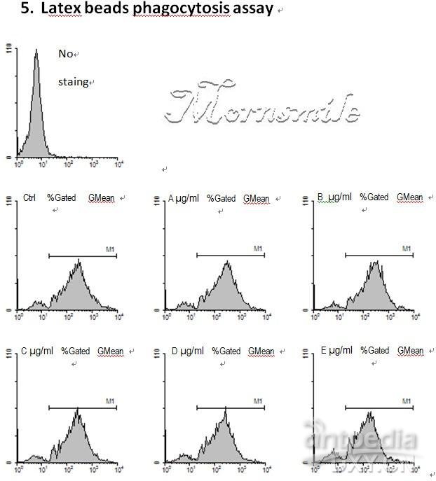
下图是专职吞噬细胞(microglia)对荧光微球的吞噬。
可见所给处理对吞噬功能无明显影响。
Histogram
很多同学都在找荧光微球体吞噬实验操作步骤,看到这篇资料非常不错,很清楚的了解微球体吞噬实验protocol,在这里分享给大家,希望对刚做荧光微球体吞噬实验的同学有帮助。