PriCells: Isolation of endothelial progenitor cells (EPCs)
PriCells: Isolation of endothelial progenitor cells (EPCs)
1. Twenty-four mL venous blood was collected at each time point into Vacutainer CPT Mononuclear Cell Preparation Tubes.
2. MNCs
recovered by density-gradient centrifugation in these tubes were washed
twice with PBS and once in EPC growth media consisting of medium199
supplemented with 20% fetal bovine serum, penicillin (100 U/mL), and
streptomycin (100 μg/mL).
3. Cells were resuspended in media, plated at a density of 5x10⁶ per well on dishes coated with human fibronectin, and incubated at 37°C in humidified 5% CO2.
4. After
48 hours, nonadherent cells suspended in the growth media were replated
onto fibronectin-coated 24-well plates at a density of 106 per well.
5. Media was changed every 3 days.
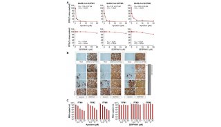
EPC colony-forming units after 7 days in culture (Figure A).
Cell clusters alone without emerging spindle cells (Figure B).
Reference
Tiffany M. Powell, Jonathan D. Paul,
Jonathan M. Hill, Michael Thompson, Moshe Benjamin, Maria Rodrigo, J.
Philip McCoy, Elizabeth J. Read, Hanh M. Khuu, Susan F. Leitman, Toren
Finkel, Richard O. Cannon III. Granulocyte Colony-Stimulating Factor
Mobilizes Functional Endothelial Progenitor Cells in Patients With
Coronary Artery Disease. Arteriosclerosis, Thrombosis, and Vascular
Biology. 2005; 25: 296-301
-
科技前沿

-
科技前沿

-
焦点事件


