卵巢癌诊断新工具——光声成像
光声成像新突破——
光声成像检测卵巢癌
关键词:光声成像; 拉曼共振吸收; SERRS; 表面增强拉曼光谱法; 金纳米棒; 卵巢癌; Endra nexus 128
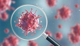
卵巢癌是女性生殖器官常见的肿瘤之一,发病率仅次于子宫颈癌和子宫体癌而列居第三位。但因卵巢癌致死者,却占各类妇科肿瘤的首位,对妇女生命造成严重威胁。国内外临床资料统计,如能得到早期诊断,90%的病人通过治疗都能活下来;但如果发展到晚期,癌细胞扩散到卵巢,存活率将低于30%。由于卵巢癌临床症状隐匿,绝大多数患者就诊时已是临床晚期。因此早期发现、早期诊断是提高治愈率、延长生存期和改善生存质量的前提和保障。
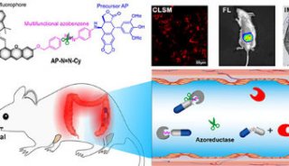
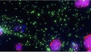
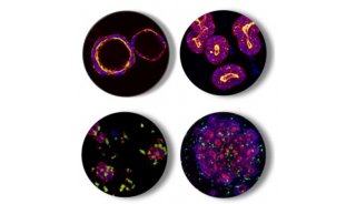
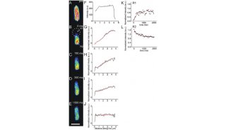
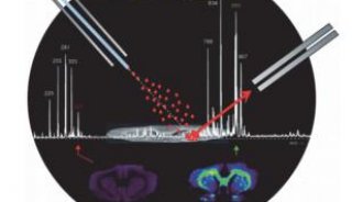
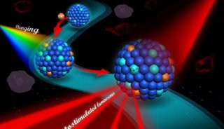
近来,斯坦福大学的研究者们首次通过光声成像和拉曼成像,以金纳米棒为肿瘤靶向标记物,观察到活体小鼠皮下移植的2008, HEY, 和SKOV33种细胞系卵巢肿瘤。研究发现,金纳米棒注入小鼠体内3小时即可观察到最大强度的光声信号,信号可以一直持续到注射后两天;金纳米棒浓度越高,光声信号越强,二者有良好的线性关系。通过拉曼成像,还可以清晰的区分肿瘤边缘与正常组织。首次使用光声和拉曼造影剂实现活体内卵巢癌成像。

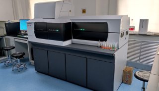
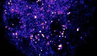
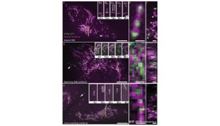
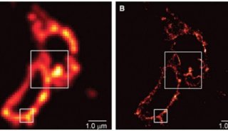
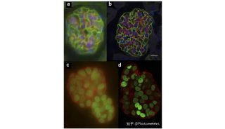
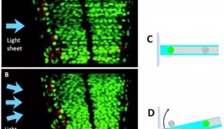
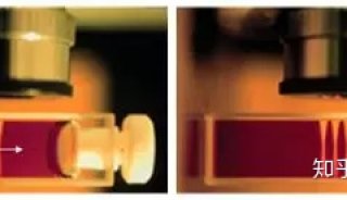
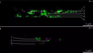
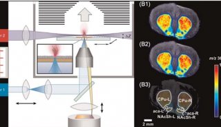
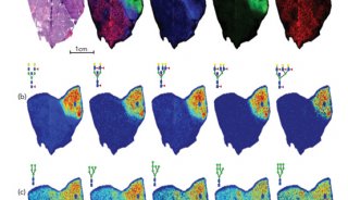
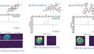
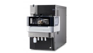
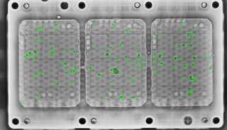
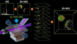
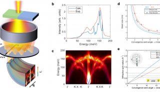
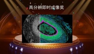
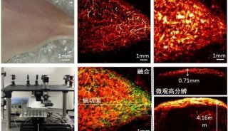
利用金纳米棒对MDA-435S(A,对照组) 2008(B), HEY(C), 和SKOV3(D)卵巢肿瘤的光声成像(仪器:Endra Inc.Nexus 128)

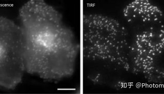
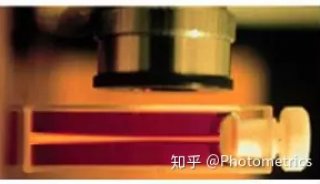
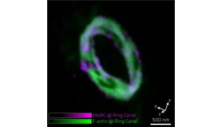
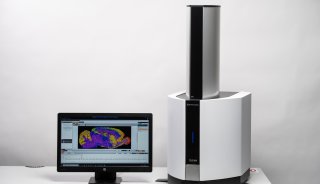
卵巢癌的拉曼成像。M:正常组织T:肿瘤(仪器:InVia, Renishaw)
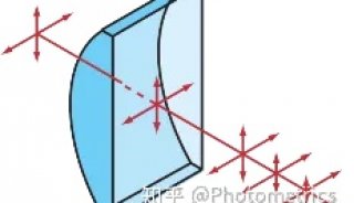
金纳米棒在近红外波段对光有强烈的散射,而生物体在这个波段的散射背景较弱,使得金纳米棒可以作为自由出入人体组织光学窗口的近红外荧光探针,经过多种基团修饰后获得对肿瘤细胞的靶向性。金纳米棒通过可调节的长径比,其等离子共振吸收峰会产生不同程度的红移。本文研究了3种长径比的金纳米棒,最终选取其共振吸收波峰750nm的作为光声成像和拉曼成像两种成像模式的造影剂,对卵巢癌取得较好的观察效果。
这些研究成果为金纳米棒的肿瘤早期探测、分子影像与靶向治疗等方面的潜在应用打下了良好基础。目前卵巢癌的早期诊断方法主要是肿瘤标记物和现代影像学检查。影像学主要包括CT、MRI、PET等手段。其中B超以实时、费用较低的特点,是首选检测工具。B超辅以光声成像有望为肿瘤临床检测提供更有利的证据。
光声成像等先进的成像技术将为肿瘤的临床早期检测提供更可靠、准确的诊断手段。
原文摘要:
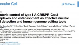
<Gold Nanorods for Ovarian CancerDetection with Photoacoustic Imaging and Resection Guidance via Raman Imaging in Living Mice>
†Molecular Imaging Program at Stanford (MIPS), Department of Radiology, Stanford University, 318 Campus Drive, Stanford, California 94305-5427, United States,
and ‡Bioengineering, Materials Science & Engineering, Bio-X, Stanford University, Stanford, California 94305, United States
Abstract:Improved imaging approaches are needed for ovarian cancer screening, diagnosis, staging, and resection guidance. Here, we propose a combined photoacoustic (PA)/Raman approach using gold nanorods (GNRs) as a passively targeted molecular imaging agent. GNRs with three different aspect ratios were studied.Those with an aspect ratio of 3.5 were selected for their highest ex vivo and in vivo PA signal and used to image subcutaneous xenografts of the 2008, HEY, and SKOV3 ovarian cancer cell lines in living mice. Maximum PA signal was observed within 3 h for all three lines tested and increased signal persisted for at least two days postadministration. There was a linear relationship (R2 = 0.95) between the PA signal and the concentration of injected molecular imaging agent with a calculated limit of detection of 0.40 nM GNRs in the 2008 cell line. The same molecular imaging agent could be used for clear visualization of the margin between tumor and normal tissue and tumor debulking via surface-enhanced Raman spectroscopy (SERS) imaging. Finally, we validated the imaging findings with biodistribution data and elemental analysis. To the best of our knowledge, this is the first report of in vivo imaging of ovarian cancer tumors with a photoacoustic and Raman imaging agent.
-
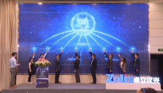
企业风采
-
企业风采
-
会议会展
-
企业风采
-
科技前沿

-
焦点事件
-
焦点事件

-
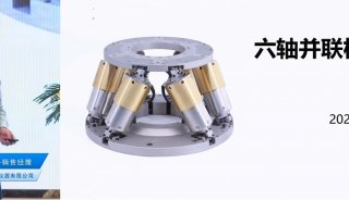
技术原理
-
科技前沿

-
招标采购

-
项目成果
-
焦点事件
-
项目成果

-
项目成果
-
市场商机
-
项目成果
-
项目成果
-
焦点事件

-
企业风采
-
科技前沿
-
科技前沿
-
精英视角
-
科技前沿

-
焦点事件
-
焦点事件

-
科技前沿
-
企业风采

-
技术原理
-
项目成果
-
焦点事件

-
企业风采
-
焦点事件

-
焦点事件

-
焦点事件

-
企业风采

-
企业风采

-
焦点事件

-
焦点事件

-
焦点事件

-
项目成果
-
精英视角

-
焦点事件

-
科技前沿

-
焦点事件

-
焦点事件

-
焦点事件

-
焦点事件

-
焦点事件

-
焦点事件

-
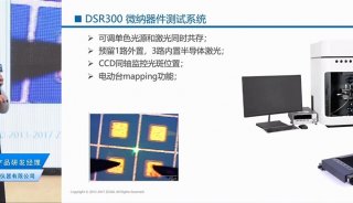
科技前沿
-
焦点事件
-
项目成果
-
焦点事件

-
焦点事件

-
焦点事件
-
项目成果
-
项目成果
-
项目成果
-
精英视角
-
焦点事件
-
项目成果

-
科技前沿
-
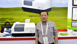
产品技术

-
焦点事件

-
科技前沿

-
项目成果
-
焦点事件

-
项目成果