激光扫描影像系统在3D肿瘤球体或干细胞克隆球体快速...2
结果:
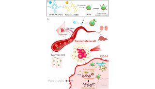
肿瘤微球体生长在超低粘附板中
|
A 96孔板 |
|
 |
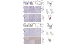
Figure 2A: 生长在96孔超低粘附板中的HepG2肿瘤微球体的全孔TIFF图像,图像底部显示了加入每孔的阿霉素浓度。2B-C:药物量效曲线(mean±SEM,n=6) |
|
B |
C |
 |
 |
|
A 384孔板 |
|
 |
Figure 3A:生长在384孔超低粘附板中的HepG2肿瘤微球体的全孔TIFF图像,图像底部显示了加入每孔的阿霉素浓度。3B-C:药物量效曲线(mean±SEM,n=8) |
|
B |
C |
 |
 |
肿瘤微球体生长在软琼脂中
|
A |
B |
C |
 |
 |
|
|
D |
||
 |
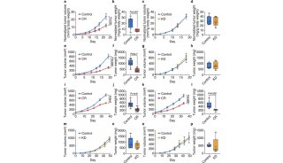
Figure 4A: 生长在96孔板软琼脂中的HepG2肿瘤微球体的全孔TIFF图像,图像上显示了加入每孔的阿霉素浓度。4B-D:药物量效曲线(mean±SEM,n=3) |
|
A |
B |
C |
 |
 |
|
|
D |
||
 |
Figure 5A: 生长在384孔板软琼脂中的HepG2肿瘤微球体的全孔TIFF图像,图像上显示了加入每孔的阿霉素浓度。5B-D:药物量效曲线(mean±SEM,n=4) |
总结:
生长在 3D培养系统中的细胞群体、不同细胞类型、微组织结构相互协调、相互作用,模拟了生命物质在体内微环境中的真实生存环境,为研究者对疾病的剖析和攻克提供了高质量的数据信息。我们在96和384微孔板中建立了两种简单且稳定的进行肿瘤微球体3D培养的方法,通过染色和图像采集,获得数目、面积、体积和荧光强度等信息。药物量效曲线的一致性结果体现了两种方法的可靠性和可重复性,这些数据表明acumen hci激光扫描系统是对基于两种培养系统获得的肿瘤微球体进行高通量(5分钟/板)分析的理想平台,该平台在过去的研究中已经被广泛地应用于肿瘤学[7-9]和干细胞研究领域[10-12]。
References:
1. Maria V, Sharon G, Frances B, et al. Advances in establishment and analysis of three dimensional tumor spheroid-based functional assays for target validation and drug evaluation. BMC Biology, 2012, 10:29
2. Patricio G, Nicola J H, Ute A, et al. Recent advances in 2D and 3D in vitro systems using primary hepatocytes, alternative hepatocyte sources and non-parenchymal liver cells and their use in investigating mechanisms of hepatotoxicity, cell signaling and ADME. Arch. Toxicol, 2013, 87(8): 1315-1530
3. Yinzhi L, Amish A, Ke Ch, et al. Neural Cell 3D Microtissue Formation is Marked by Cytokines’ Up-Regulation. PLoS One, 2011, 6(10): e26821
4. Yesl J, Ah R K, Jae S L. 3D co-culturing model of primary pancreatic islets and hepatocytes in hybrid spheroid to overcome pancreatic cell shortage. Biomaterials, 2013, 34(15): 3784-3794
5. Anna I A, Brenda K M, Glen D P, et al. A 3-D organoid kidney culture model engineered for high-throughput nephrotoxicity assays. Biomaterials, 2012, 33(18): 4700-4711
6. Sasai Y. Next-generation regenerative medicine: organogenesis from stem cells in 3D culture. Cell Stem Cell, 2013, 12(5): 520-530
7. Weijuan W, Chen Bi, Kelly M C, et al. Inhibition of tumor growth and metastasis in non-small cell lung cancer by LY2801653, an inhibitor of several oncokinases, including MET. Clin Cancer Res, 2013, 19(20): 5699–5710
8. Kai W, Ho Y L, Shephanie S, et al. Genomic Landscape of Copy Number Aberrations Enables the Identification of Oncogenic Drivers in Hepatocellular Carcinoma. Hepatology, 2013, 58(2): 706-717
9. Shane R H, Jeremy T, Anthony P O, et al. An HTS-Compatible 3D Colony Formation Assay to Identify Tumor-Specific Chemotherapeutics. J Biomol Screening, 2013, 18(10): 1298-1308
10. Koppany V, Hydeyuki O, Robert D, et al. A molecular screening approach to identify and characterize Inhibitors of glioblastoma stem cells. Mol Cancer Ther, 10(10), 1818-1828
11. Anne D, Jimmy E, Richard J S, et al. CXCR4 Expression in Prostate Cancer Progenitor Cells. PLoS One, 2012, 7(2): e31226
12. Claudia P, Ina K, Leoni K. Discovery of the cancer stem cell related determinants of radioresistance. Radiother Oncol, 2013, 108(3), 378-387
-
焦点事件
-
招标采购

-
科技前沿
-
企业风采

-
科技前沿

-
科技前沿
-
焦点事件

-
项目成果
-
招标采购

-
项目成果

-
企业风采
-
产品技术
-
科技前沿

-
精英视角
-
科技前沿

-
科技前沿
-
焦点事件

-
技术原理

-
焦点事件

-
技术原理
-
焦点事件
-
产品技术

-
科技前沿
-
项目成果

-
焦点事件

-
产品技术
-
产品技术

-
焦点事件

-
焦点事件

-
焦点事件

-
项目成果

-
项目成果
-
综述
-
企业风采
-
项目成果