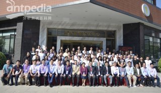
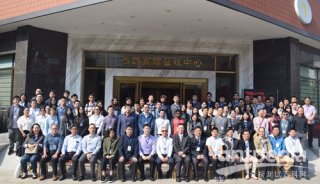
第八届全国微全分析系统学术会议微纳米生物分析专场
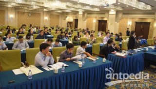
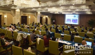
2013年5月16日-19日,由中国化学会主办、厦门大学承办、复旦大学、浙江大学协办的为期四天的第八届全国微全分析系统学术会议、第三届全国微纳尺度生物分离分析学术会议暨第五届国际微化学与微系统学术会议在美丽的海滨城市厦门隆重召开。以下是5月17日微纳米生物分析专场报告。
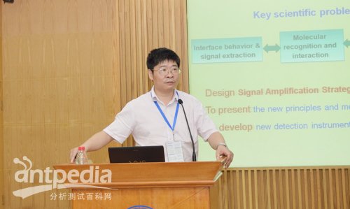
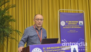
首先来自南京大学的鞠熀先教授为我们带来了题为《Signal amplification coupled with molecular recognition for biological analysis》的精彩报告,以下是摘要原文:
This talk will introduce a series of novel signal amplification strategies and their application in biosensing and biological analysis based on nanotechnology and molecular biological methods. The nanotechnology for signal amplification includes:
1) accelerating the electron transfer or obtaining sensitized optical signal, 2) catalyticand enzyme mimetic functions of the nanomaterials, 3) using nanomaterials as tag molecules, 4) using nanomaterials as the carriers of signaling molecules, 5) electrochemiluminescent or photoelectrochemical signal amplification, and 6) selective concentration of biomolecules by biorecognition. The molecular biological amplification is designed by introducing rolling circle amplification, target-induced repeated primer extension, hybridization chain reaction, loop-mediated amplification, target DNA recycling amplification such as endonuclease-, exonuclease- and polymerase-based circular strand replacement polymerization to amplify the electrochemical, optical and visual signals. The established methods can conveniently be used in the detections of small biomolecules, proteins, cells, the carbohydrate sites on cell surfaces and intracellular microRNA by electrochemical, optical, mass spectrometric, and imaging measurements.

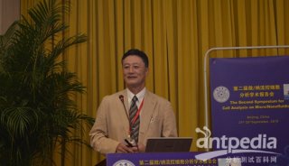
来自厦门大学的任斌教授为我们带来了题为《Plasmon-enhanced Raman Spectroscopy for Surface- and Bio-analysis》的精彩报告。以下是内容摘要。
Both surface-enhanced Raman spectroscopy (SERS) and tip-enhanced Raman spectroscopy (TERS) is benefited from the the localized surface plasmon resonance (LSPR) of metallic nanostructures. They can provide high sensitivity with molecular fingerprint information for ultratrace analysis, even down to single molecules. To take the challenge of using SERS for bioanalysis , we developed a method to modify the surface of SERS active nanoparticle colloids or solid SERS substrates with some halide ions. Proteins were found to interact with the modified substrate via electrostatic interaction. The SERS signal of protein is at least enhanced by 1000 time over the solution species, with almost identical feature to that of the solution signal of proteins. The methods have been applied to study the lysosome, BAS, avidin, hemoglobin, cytochrome c and etc. The detection limit for lysosome can be as low as 3 mg/mL. The ability to obtain SERS signal of protein with very good reproducibility and high sensitivity is extremely important to the wider application of SERS technique to biological systems. We further systematically study the methodology of using a probe molecule to monitor the local pH environment of live cells. It was found that it is vitally important to control the interfacial structure and measuring condition in order to obtain reliable pH response. On the other hand, TERS can not only provide very high sensitivity but also high spatial resolution, which is extremely important when it is used to study the dynamic processes on surfaces. The high spatial resolution allows the extraction of signals from the “some molecules” or even “single molecules” by significantly lowering the background averaged signal. Using thiols for example, we found that TERS can clearly provide the immersing time dependent of the self-assemble mononlayer, which is a reflection of the strong interaction between the thiol molecules. The dynamic diffusion process could be revealed by the combined two-dimensional and autocorrelation analyses.

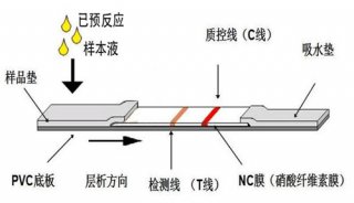
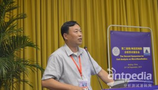
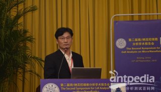
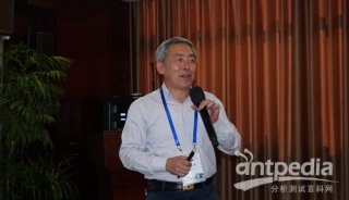
来自南京大学化学化工学院的许丹科教授为我们带来了题为《基于纳米银探针的微阵列芯片检测方法的研究》的精彩报告,以下是内容摘要:
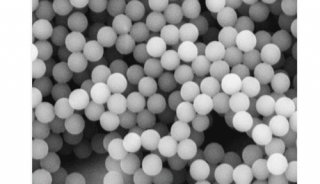
纳米材料的功能化研究对于发展其在生物分析领域的应用具有重要作用。近年来,我们研究了纳米银的合成及功能化方法。通过巯基自组装、亲和素-生物素相互作用等途径实现了纳米银表面的DNA 单链分子、适配体及蛋白质分子等的功能化修饰,采用研制的功能化纳米银分析材料建立了电化学检测、荧光检测及可视化检测等多种微阵列芯片的分析方法。
以DNA 修饰的纳米银为生物功能探针、DNA 芯片为分析模式,开展了对多种病毒DNA片段的电化学信号放大检测方法的研究,具有快速简便、灵敏度高、可实现多靶标检测等优点。开展了功能化纳米银荧光增强检测方法的研究,研究了基于纳米银荧光增强机制的新型生物探针及基于聚合纳米银的金属荧光增强检测方法,利用Cy3 分子数量的增加及Cy3 发射光谱与纳米银聚合物表面等离子体共振峰的耦合作用放大荧光信号,实现对蛋白质的高灵敏检测。此外,研究了适配体修饰的可视化纳米银生物探针检测方法,将微孔板生物芯片技术与纳米银催化银离子显色方法结合,具有灵敏度高、操作简单、结果可视化的优点。
研究内容揭示了功能化纳米银分析材料具有优良的生物兼容性,能成为DNA、蛋白质等多种生物分子识别的有效载体。纳米银本身显示了良好的光电特性,为发展新型纳米分析材料提供了新的发展方向。
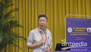
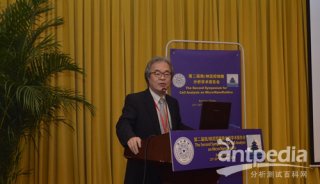
来自材料研究工程学院的苏晓迪为我们带来了题为《Hybrid Assembly of Gold Nanoparticles with Fluorescent Materials for Studying Protein-DNA Interaction and Ligand Inhibition》
Gold nanoparticles have unique optical properties arising from their ability to support localized surface plasmonc resonance. Gold nanoparticles are also well-known by their ability to alter the emission properties of proximal fluorophores, due to Förster resonance energy transfer (FRET), nanomaterial surface energy transfer (NSET), or electron transfer, depending on the distance of the fluorophores to nanoparticle surface and the emission wavelength of the fluorophores. In previous studies, we have developed a series of metal nanoparticles based bioassays for studying various biomolecular binding events, concerning DNA mutation [1-3], gene transcription [4, 5], enzymatic cleavage of DNA, and aptamer selection [7], by exploiting interparticle distance determined plasmonic property. In this study we have combined gold nanoparticles’ fluorescence quenching properties and protein-DNA binding induced hybrid assembly between gold nanoparticles and fluorescent materials for efficient study of transcription factor-DNA interactions and ligand inhibition that are important for breast cancer research and drug discovery. The results are compared with those using conventional biological analysis methods, i.e. EMSA and fluorescence anisotropy, as well as instrument based methods (dual polarization interferometer and surface plasmon resonance spectroscopy). With the conventional methods as reference, we concluded that the gold nanoparticle-fluorophore hybrid sensors have higher sensitivity to determine subtle affinity difference induced by single base mutation in DNA elements and to report strong and weak ligand interruption. Understanding the DNA binding property and ligand effects of these transcription factors is of significant for breast cancer and drug discovery research.
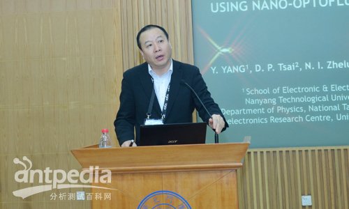
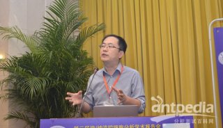
来自南洋理工大学的杨毅教授为我们带来了题为《Microparticles Sorting by Hydrodynamic Optical Forces》的精彩报告,以下是报告摘要:
This paper reports a microfluidic microparticles sorting generator by using the optical force and hydrodynamic forces. Micro/Nanoparticles ranging in size from 70 nm to 1 μm can be aligned and focused in the core flow stream by hydrodynamic focusing [1-3]. The particles can be subsequently manipulated by the optical force and liquid stoke forces. The particles with different size and refractive indices can be selected by the optical forces and drag force, flowing with different traces. It has a great potential in cell, or molecule sorting and Separating.
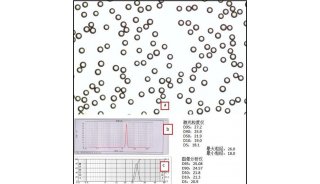
Figure 1 shows the schematic of the nano-optofluidic system, which consists of three flow streams in a microchannel. Nanoparticles are flowing in the core flow stream by the hydrodynamic effect. A single-mode optical fiber is injected vertically. When the particles flow in the microchannel, they will be controlled by three forces: the optical gradient force that pushes particles to the higher intensity, the radiation force that exists in the direction of light propagation, and the drag force that hinders the velocity change of the particles. In principle, larger particles are more easily to be moved by optical forces while smaller particles are easily blocked by the drag force due to its smaller specific surface area. As a result, particles with different sizes can be sorted by a suitable flow rate and input power in the microchannel.
Figure 2 shows that optical forces are very sensitive to the refractive indices and extinction coefficient of particles. Fig. 3 shows the simulation results of the nanoparticles under optical field in the central of the input power. Fig. 4 shows the images of the sorting traces of different size particles in the microchannel under the optofluidic forces with different diameters.
In conclusion, an optofluidic device for single cell and molecule sorting by using optical force combined with the hydrodynamic focusing is demonstrated.
-
标准
-
投融资
-
企业风采

-
精英视角
-
企业风采
-
项目成果

-
并购

-
企业风采

-
企业风采
-
企业风采

-
投融资
-
焦点事件

-
焦点事件

-
企业风采

-
精英视角

-
科技前沿
-
焦点事件

-
焦点事件

-
焦点事件
-
技术原理